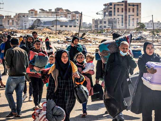
Buonasera a tutti e a tutte, ci eravamo abituati, assuefatti a continuare a negare, a rigettare tutto quello che abbiamo passato ognuno di noi nel periodo 2020-2021 e nel 2022-2023, ma oggi le circostanziali parole di Giada Borgato, la ciclista giornalista sportiva Rai che seguiva i ciclisti del gruppo nella tappa numero 11, con partenza da Fojano di Val Fortore con arrivo a Francavilla al Mare in Abruzzo, hanno fatto riemrgere i dubbi a molte persone sul fatto che il pericolo da virus sia ancora aleggiante tra noi. Il presente articolo servirà a fare luce sulla reale causa di ritiri multipli dei ciclisti al Giro d’Italia: durante la prima settimana di manifestazione se ne sono ritirati ben 13, oggi altri quattro e durante le telecronache su RaiDue erogate dal servizio pubblico televisivo, rappresentato da Rai RadioTelevisione Italiana, cerca continuamente e goffamente di nascondere in tutti i modi che si tratta di COVID19, non chiamando più il virus in tal modo, ma etichettandolo come un semplice virus intestinale che però guarda caso sta facendo tornare le broncopolmoniti in gruppo, pertanto il virus in questione non è classificabile come un lieve virus intestinale e si deve continuare ad avere il coraggio di chiamarlo con il nome con cui lo abbiamo menzionato per quattro anni e dovremmo farlo ancora per diverso tempo. Io dico sempre che i ciclisti amatoriali non devono mai emulare i professionisti, anche quando ci si prende una malattia subdola e meschina qual’è il COVID19, perché la differenza tra ciclisti professionisti e ciclisti amatori è presto detta: loro sono seguiti e possono avere accesso a cure costose ed eccelse, come anticorpi monoclonali e farmaci antivirali non disponibili nelle nostre comuni farmacie, cure alle quali un comune proletario mortale non può assolutamente accedere, dovendosi poi curare da solo ed empiricamente con antibiotici ed anti infiammatori e con un Sistema Sanitario Nazionale (SSN) in costante decadenza, per cui continuare a vedere in giro amaori in gruppo che si comportano esattamente come i professionisti, è soltanto follia pura e ve lo scrive un ciclista amatoriale che è andato in bicicletta da corsa dal 2008 al 2019, quindi non sono proprio il primo stronzo venuto che parla a struso, io parlo e scrivo sempre con “cognitionis causae”! Tutti questi indegni miseri tentativi sionisti di non rivelare la verità rischiano di far perdere di credibilità il servizio pubblico italiano televisivo e nonostante tutta la dittatura sionista di regime dalla quale siamo continuamente bombardati tutti i giorni attraverso i principali media di Stato, il mio interesse verso il Giro d’Italia non cesserà mai, perché la passione del ciclismo si trasmette anche attraverso immagini televisive e non soltanto praticando uno degli sport più interessanti ed appassionanti al Mondo e per quanti miseri tentativi si continueranno a portare avanti per mistificare la realtà e perseguire gli interessi del sionismo, che ha pianificato un deliberato attentato criminale alla salute di ognuno di noi nel Marzo 2020 e che continua a non cessare, il potere illuminante della verità un giorno farà emergere tutta questa machiavellica messa in scena, atta a trasformare l’Umanità in una società Post-Umanista al servizio del profitto delle lobby e multinazionali sioniste ed io con una società occidentale decadente del genere, non voglio avere mai più niente a che fare, perché almeno lo sport che dovrebbe garantire libertà di espressione, oggi non è più credibile e deve tornare ad avere quella credibilità che merita, a prescindere dagli interessi economici di palazzo che deve conseguire il sionismo!
Viva il Giro d’Italia, viva laverità anti-sionista e viva la Palestina Libera!
THE WITHDRAWALS AT THE 2024 GIRO D’ITALIA DUE TO A “MYSTERIOUS INTESTINAL VIRUS”: YOU NO LONGER HAVE THE COURAGE TO CALL IT COVID19!
Good evening to everyone, we had become accustomed, accustomed to continuing to deny, to reject everything that each of us went through in the period 2020-2021 and 2022-2023, but today the circumstantial words of Giada Borgato, the cyclist sports journalist Rai who followed the cyclists of the group in stage number 11, starting from Fojano di Val Fortore and arriving in Francavilla al Mare in Abruzzo, raised many people’s doubts about whether the danger from the virus is still hovering among us. This article will serve to shed light on the real cause of multiple withdrawals of cyclists from the Giro d’Italia: during the first week of the event 13 withdrew, today another four and during the commentary on RaiDue provided by the public television service, represented from Rai RadioTelevisione Italiana, continually and clumsily tries to hide in every way that it is COVID19, no longer calling the virus in this way, but labeling it as a simple intestinal virus which, however, coincidentally is causing bronchopneumonia to return in groups, therefore the virus in question cannot be classified as a mild intestinal virus and we must continue to have the courage to call it by the name with which we have mentioned it for four years and we should do so for some time to come. I always say that amateur cyclists must never emulate professionals, even when you catch a subtle and petty disease such as COVID19, because the difference between professional cyclists and amateur cyclists is easy to tell: they are followed and can have access to expensive and sublime treatments, such as monoclonal antibodies and antiviral drugs not available in our common pharmacies, treatments to which a common mortal proletarian absolutely cannot access, having to then treat himself alone and empirically with antibiotics and anti-inflammatories and with a National Health System ( SSN) in constant decline, so continuing to see amaori around in groups who behave exactly like professionals is just pure madness and this is written to you by an amateur cyclist who rode a racing bicycle from 2008 to 2019, so they are not the first asshole to come and talk nonsense, I always speak and write with “cognitionis causae”! All these unworthy miserable Zionist attempts not to revealing the truth risks causing the Italian public television service to lose credibility and despite all the Zionist dictatorship by which we are continuously bombarded every day through the main state media, my interest to watch the Giro d’Italia will never cease, because the passion for cycling is also transmitted through television images and not only by practicing one of the most interesting and exciting sports in the World and no matter how many miserable attempts will continue to be made to mystify reality and pursue the interests of Zionism, which planned a deliberate criminal attack on the health of each of us in March 2020 and which continues to continue, the illuminating power of the truth one day will bring out all this machiavellian staging aimed at transforming Humanity into a Post-Humanist society at the service of the profit of Zionist lobbies and multinationals and I never want to have anything to do with a decadent Western society like that again, because at least sport, which should guarantee freedom of expression, is no longer credible today and must return to having the credibility it deserves, regardless of the economic interests of the palace that Zionism must achieve!
Long live the Giro d’Italia, long live the anti-Zionist truth and long live Free Palestine!
Prima della partenza di questa tappa si sono ritirati altri quattro corridori più i 13 della fine della scorsa settimana causa sconosciuta, ma #GiadaBorgato rivela una causa che non si vuole ammettere per non dare allarmismo: "#Febbre alta da #virus e #broncopolmoniti" in gruppo.
Before the start of this stage, another four riders withdrew plus the 13 at the end of last week due to an unknown cause, but #GiadaBorgato reveals a cause that doesn't want to admit in order not to cause alarmism: "High #fever from a #virus and #bronchopneumonia" in group.
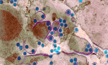
Il #virus in questione di cui ha parlato #GiadaBorgato durante la diretta della tappa su #Raidue è il #COVID19 che continua a girare tra noi, ma nessuno ha il coraggio di ammettere pur di "doverci convivere" come hanno stabilito gli stessi #sionisti che bombardano #Gaza!
The #virus in question that #GiadaBorgato talked about during the stage broadcast on #Raidue is the #COVID19 that continues to circulate among us, but no one has the courage to admit in order to "have to live with it" as the #Zionists themselves have established who bomb #Gaza!
Dott. Alessio Brancaccio, tecnico ambientale Università degli Studi di L’Aquila, membro della Fondazione Michele Scarponi Onlus, ideologo e membro del movimento ambientalista Ultima Generazione A22 Network per contrastare il Riscaldamento Globale indotto artificialmente dalla Geoingegneria Solare SRM
Clementi fa il punto su strumenti che abbiamo e potremo avere, ‘quando suona allarme cruciale saper rispondere’
Milano, 3 mag. (Adnkronos Salute) – Da inizio aprile gli Usa, dopo il rilevamento dell’influenza aviaria A H5N1 ad alta patogenicità negli allevamenti di bovini da latte di alcuni Stati e un collegato caso umano (un lavoratore del settore lattiero-caseario in Texas), si interrogano sul rischio di ulteriori sviluppi della situazione. Si valutano gli scenari peggiori: che il virus si evolva ancora, diventando in grado di trasmettersi da uomo a uomo. La domanda ricorrente: il Paese sarebbe pronto per una pandemia? Perché lo sia si lavora a strategie e si fa l’inventario di quali vaccini e armi terapeutiche potranno essere necessari. E l’Italia come deve muoversi? Che strumenti servono per prepararsi a fronteggiare eventuali rischi? “Antivirali, vaccini”, ma non solo, spiega all’Adnkronos Salute il virologo Massimo Clementi. Parola chiave: “Vigile attesa”, dice l’esperto.
“Ad oggi – fa il punto – per quanto riguarda gli antivirali abbiamo farmaci che bloccano una delle due proteine del virus, la neuraminidasi, che sono piuttosto efficaci, anche se ovviamente entrano in gioco nel momento in cui è già iniziata l’infezione. Di preventivo c’è ovviamente il vaccino ed eventualmente anticorpi monoclonali. Questi ultimi però devono essere anticorpi monoclonali che riconoscono l’emoagglutinina” presente sulla superficie “di questo virus”. In ogni caso, “abbiamo alcuni presidi piuttosto efficaci, ma bisogna conoscere il virus”. E poi “c’è la possibilità di avere diverse tipologie di vaccini: il vaccino classico che si ottiene in uova embrionate, è mediamente efficace ed è come quello che abbiamo utilizzato ogni anno per proteggerci nei confronti dell’influenza; più efficaci e con meno effetti collaterali sono i vaccini che sono costituiti da particelle di queste proteine, assorbite su piccole vescicole di grasso, che immunizzano il soggetto che li riceve. C’è infine sempre la possibilità, così come avvenuto per Sars-CoV-2, di sviluppare vaccini di diversa natura che però in questo momento non ci sono”.
Ma per l’Italia, come per altri Paesi, “la cosa più importante in assoluto è una strategia di controllo – puntualizza Clementi – per verificare in primo luogo che cosa sta succedendo nelle specie selvatiche che arrivano da noi e non possono essere bloccate. Serve per questo monitorare, e lo fa egregiamente nel nostro Paese l’Istituto zooprofilattico delle Venezie. E poi è anche da valutare che cosa accade negli allevamenti di diverse specie, ma in questo momento c’è veramente un monitoraggio molto stretto su questo, da quelli di animali allevati per scopi alimentari fino a quelli che lo sono per scopi diversi, come visoni, marmotte e così via. Ma io su questo piano mi sento molto tutelato dalla struttura e dalla rete efficace che il nostro Paese ha, forse migliore di altri in questo caso. Certo, quando suona l’allarme bisogna essere in grado di rispondere a quell’allarme”.
“Insomma – ripete il virologo, che per anni ha diretto il Laboratorio di microbiologia e virologia dell’ospedale San Raffaele di Milano – al momento la situazione deve essere di vigile attesa, non c’è oggi niente di clamorosamente pericoloso. Però queste mie parole domattina potrebbero essere smentite da un nuovo evento che cambia il quadro”, avverte. “Come detto più volte, il fatto che ci sia stata una persona infettata in un allevamento non rappresenta in sé un fatto estremamente pericoloso”, perché non è sinonimo di trasmissione uomo-uomo. “Rappresenta chiaramente un evento su cui porre attenzione”.
Quello che sarebbe da considerare pericoloso, conclude, “è il fatto che il virus possa diventare umano. C’è un’altra possibilità: che il virus aviario mescoli il proprio genoma con altri virus influenzali, come è successo non tanti anni fa con un virus del maiale che si era mescolato geneticamente e aveva dato luogo a un nuovo virus capace di infettare l’uomo. Tanto che era stato avviato un allarme per una pandemia che poi è rientrato, perché in realtà si trattava di infezioni di modesta entità clinica”.
Fonte: La Gazzetta del Mezzogiorno
English translate
BIRD FLU: ‘ANTIVIRALS, VACCINES AND WATCHING WAITING’, HOW WE PREPARE FOR THE RISK OF A SECOND PANDEMIC
Clementi takes stock of the tools we have and could have, ‘when an alarm goes off it is crucial to know how to respond’
FRIDAY 03 MAY 2024, 3.50pm
Milan, 3 May. (Adnkronos Health) – Since the beginning of April, the USA, after the detection of highly pathogenic avian influenza A H5N1 in dairy cattle farms in some states and a related human case (a worker in the dairy sector in Texas), has they ask about the risk of further developments in the situation. The worst scenarios are evaluated: that the virus evolves further, becoming capable of transmitting from human to human. The recurring question: would the country be ready for a pandemic? For this to be the case, we work on strategies and take an inventory of which vaccines and therapeutic weapons may be necessary. And how should Italy move? What tools are needed to prepare to face possible risks? “Antivirals, vaccines”, but not only that, explains virologist Massimo Clementi to Adnkronos Salute. Keyword: “Watchful waiting”, says the expert.
“To date – he takes stock – as far as antivirals are concerned we have drugs that block one of the two proteins of the virus, neuraminidase, which are quite effective, even if they obviously come into play when the infection has already started. preventive there is obviously the vaccine and possibly monoclonal antibodies. The latter, however, must be monoclonal antibodies that recognize the haemagglutinin “present on the surface” of this virus. In any case, “we have some rather effective safeguards, but we need to know the virus”. And then “there is the possibility of having different types of vaccines: the classic vaccine which is obtained in embryonated eggs, is on average effective and is like the one we have used every year to protect ourselves against the flu; more effective and with less side effects are vaccines that are made up of particles of these proteins, absorbed on small fat vesicles, which immunize the subject who receives them. Finally, as happened with Sars-CoV-2, there is always the possibility of developing vaccines of a different nature which however do not exist at this moment”.
But for Italy, as for other countries, “the most important thing of all is a control strategy – Clementi points out – to verify first of all what is happening in the wild species that come to us and cannot be stopped. for this reason, the Istituto zooprophylattico delle Venezie does it very well in our country. And then it is also necessary to evaluate what happens in the farms of different species, but at the moment there is really very close monitoring on this, from those. from animals bred for food purposes to those bred for different purposes, such as minks, marmots and so on, on this level I feel very protected by the structure and effective network that our country has, perhaps better than others this case. Of course, when the alarm sounds you have to be able to respond to that alarm.”
“In short – repeats the virologist, who for years directed the microbiology and virology laboratory of the San Raffaele hospital in Milan – at the moment the situation must be one of watchful waiting, there is nothing sensationally dangerous today. But these words of mine tomorrow morning they could be contradicted by a new event that changes the picture”, he warns. “As said several times, the fact that there was an infected person on a farm does not in itself represent an extremely dangerous fact”, because it is not synonymous with human-human transmission. “It clearly represents an event worth paying attention to.”
What would be considered dangerous, he concludes, “is the fact that the virus could become human. There is another possibility: that the avian virus mixes its genome with other influenza viruses, as happened not many years ago with a pig virus that had mixed genetically and given rise to a new virus capable of infecting humans, so much so that an alarm was raised for a pandemic which was then reverted, because in reality they were infections of modest clinical entity.”
The ongoing #COVID19 pandemic wasn't enough, now we need also #avian#flu! You are starting to understand what kind of occult, sadistic and perverse people we are all in their hands. The zero social life strategy continues for me, think about saving your ass from these bastards. pic.twitter.com/e60fg4VM7o
requires a return to #organicagriculture as soon as possible, without the use of #pesticides. It's criminal #US and #chinese models of #meat food consumption, linked to the abnormal consumption of it, that has always been wrong for how they are unealthy and cruel for animals. pic.twitter.com/vHIwH2So4M
Dott. Alessio Brancaccio, tecnico ambientale Università degli Studi di L’Aquila, membro della Fondazione Michele Scarponi Onlus, ideologo e membro del movimento ambientalista Ultima Generazione A22 Network per contrastare il Riscaldamento Globale indotto artificialmente dalla Geoingegneria Solare SRM
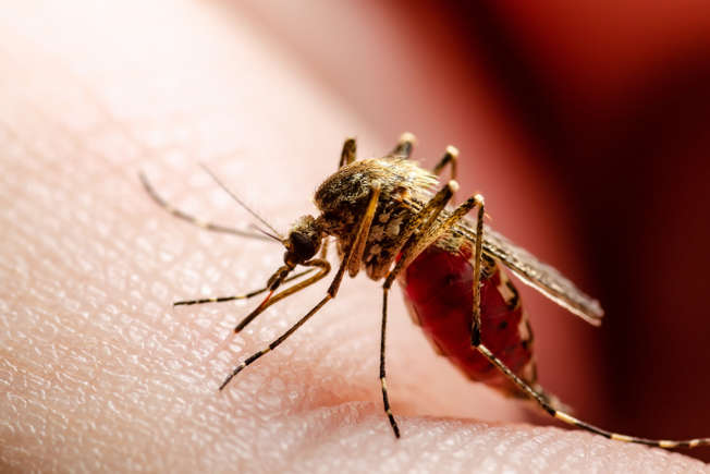
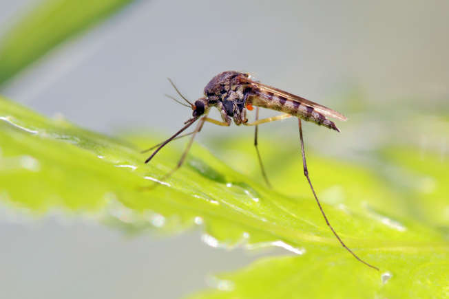
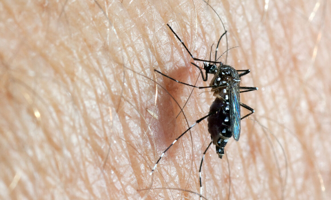
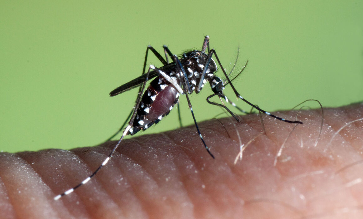
Una zanzara del genere Aedes responsabile della trasmissione del virus Dengue all’essere umano A mosquito of the Aedes genus responsible for transmitting the Dengue virus to humans
La Dengue è tornata a far parlare di sé con un aumento di casi in diversi Paesi. Porto Rico è stata particolarmente colpita e nel marzo 2024 l’isola ha dichiarato la sua prima epidemia di Dengue dal 2012. In Europa ci sono tre Paesi in cui la Dengue è approdata e l’Italia è il Paese europeo con il maggior numero di casi.
Fortunatamente la Dengue non causa malattie gravi in tutte le persone che infetta, ma i sintomi possono essere spiacevoli e a volte richiedono un intervento medico.
Cliccate sulla galleria per imparare a conoscere i sintomi della Dengue.
Virus che nasce nelle zanzare
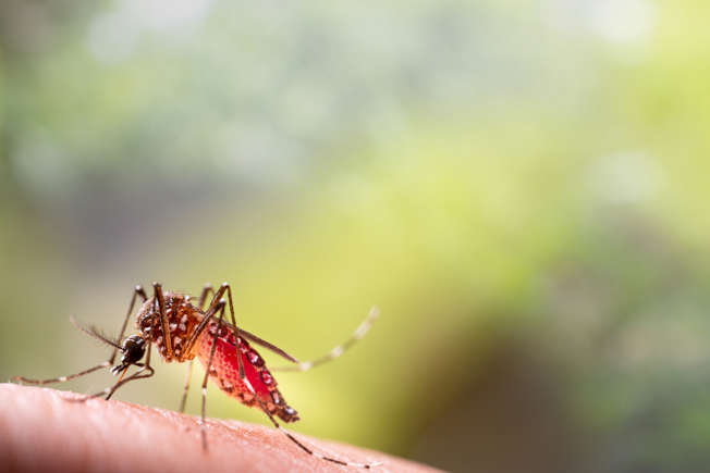
La febbre Dengue, altrimenti nota come Dengue, è un’infezione diffusa dalle zanzare. Tuttavia, non sempre provoca malattie gravi; infatti, solo una persona su quattro si ammala.
Dove è diffusa?
La Dengue è diffusa in alcune parti del mondo, tra cui alcune zone dell’Africa e dell’Asia, dell’America centrale e meridionale, dei Caraibi, delle isole del Pacifico e di alcune aree meridionali del Nord America.
È inoltre possibile contrarre la dengue in alcune zone dell’Europa meridionale in determinati periodi dell’anno (dalla primavera a Novembre).
Tra i Paesi in Europa in cui sono stati riscontrati casi di Dengue ci sono Croazia, Francia, Italia e Spagna.
Porto Rico
Sebbene la febbre dengue non sia presente ovunque nel mondo, di recente è stata al centro delle cronache per l’aumento dei casi, in particolare a Porto Rico.
Numeri
In effetti, Porto Rico ha dichiarato almeno 549 casi già nel 2024, rispetto a un numero totale di 1.293 casi per l’anno 2023.
Epidemia
Secondo il dipartimento sanitario dell’isola, più di 340 persone sono state ricoverate in ospedale a causa del virus e il Paese ha dichiarato l’epidemia.
Rischio pandemico
È la prima volta che Porto Rico dichiara un’epidemia di Dengue dal 2012, e ciò avviene dopo che l’Organizzazione Mondiale della Sanità (OMS) ha avvertito che la Dengue è un rischio pandemico nel Gennaio 2024.
Modello più ampio
In effetti, l’epidemia di Porto Rico fa parte di un modello più ampio che è emerso in tutte le Americhe fino ad ora nel 2024.
Altri Paesi
Paesi come Argentina, Brasile, Perù e Uruguay hanno riportato un numero significativo di casi di Dengue.
Riconoscere i sintomi
Anche se l’infezione da Dengue non sempre provoca la malattia, è comunque una buona idea saper riconoscere i sintomi.
Viaggiare verso un Paese dove c’è la Dengue
Questo è particolarmente vero se si intende viaggiare in un Paese in cui il numero di casi di dengue è in aumento.
Quanto ci mettono i sintomi a manifestarsi?
Se i sintomi della Dengue si manifestano, di solito si sviluppano nei 4-10 giorni successivi alla puntura di una zanzara infetta.
Sintomi simili a quelli influenzali
La febbre Dengue può sembrare un’influenza e i sintomi sono simili. Essi comprendono una temperatura elevata, un forte mal di testa e dolore dietro gli occhi.
Altri sintomi
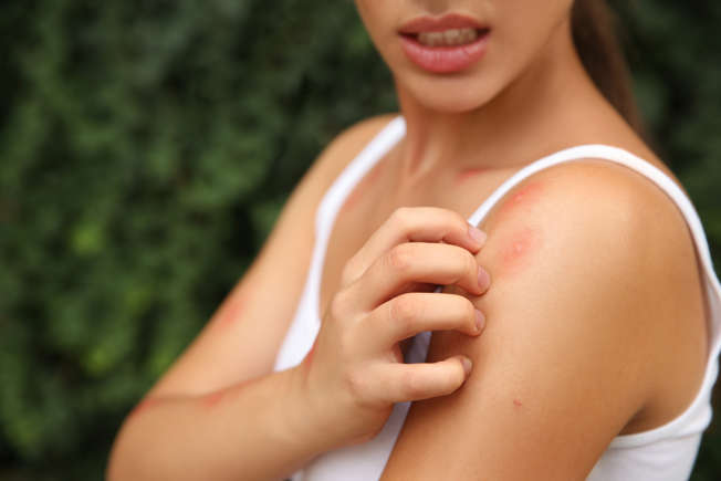
Altri sintomi includono dolori muscolari e articolari, sensazione di malessere, ghiandole gonfie e un’eruzione cutanea costituita da macchie piatte o leggermente rialzate.
Quando consultare un medico
In generale, le persone che sviluppano i sintomi della Dengue si sentono meglio dopo circa una settimana. Tuttavia, è importante rivolgersi a un medico se si è viaggiato di recente in un paese in cui è presente la Dengue e si manifestano i sintomi.
Casi di Dengue più gravi
Questo è importante perché alcune persone sviluppano un tipo di Dengue più grave pochi giorni dopo aver iniziato a sentirsi male. Si tratta di un’eventualità rara, ma non sconosciuta.
Popolazione a rischio
Chi ha già avuto la Dengue in passato ha maggiori probabilità di sviluppare una Dengue grave. È anche più comune nelle donne in gravidanza e nei neonati.
Sviluppare la Dengue con sintomi gravi
Le persone che sviluppano una Dengue grave possono iniziare a sentirsi meglio e vedere la loro temperatura tornare alla normalità, solo per sviluppare sintomi ancora più gravi 24-48 ore dopo.
Sintomi
I sintomi della Dengue grave comprendono forti dolori alla pancia, vomito ripetuto, respirazione accelerata, sanguinamento delle gengive o del naso, affaticamento, irrequietezza e vomito o feci sanguinolente.
Terapia ospedaliera
La dengue grave può diventare molto seria se non viene trattata adeguatamente in ospedale. È quindi importante rimanere vigili e rivolgersi a un medico quando necessario.
Trattamenti
Non esiste un trattamento specifico per la Dengue, ma è possibile alleviare i sintomi riposando, bevendo molti liquidi e assumendo paracetamolo.
Non assumere ibuprofene o aspirina
È importante, tuttavia, non assumere antidolorifici antinfiammatori, come l’ibuprofene o l’aspirina. Questi possono causare problemi di sanguinamento nelle persone affette da Dengue.
Evitare i viaggi
Se si appartiene a uno di questi gruppi, è consigliabile evitare di viaggiare nei Paesi in cui è presente questa infezione.
Evitare le punture di zanzara
Per le persone che viaggiano in Paesi in cui è presente la Dengue, il modo migliore per prevenire l’infezione è evitare di essere punti dalle zanzare.
Proteggersi con i vestiti
È buona norma indossare indumenti a maniche lunghe e pantaloni per coprire braccia e gambe, soprattutto nelle prime ore del mattino e della sera, quando le zanzare sono più numerose.
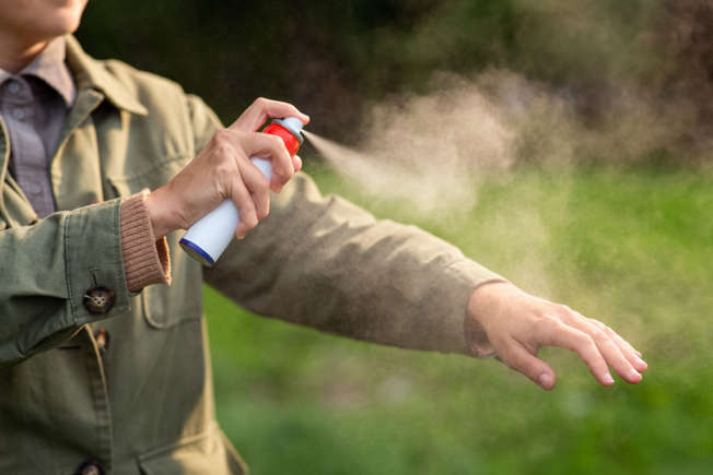
Repellente per insetti
Si può anche usare un repellente per insetti sulla pelle, preferibilmente uno che contenga DEET, e si dovrebbe cercare di chiudere le finestre e le persiane quando possibile.
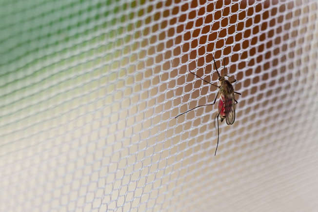
Zanzariera
Infine, è buona norma dormire sotto una zanzariera trattata con insetticida, anche quando si dorme di giorno.
In sintesi
La febbre Dengue può essere fastidiosa e i casi sono in aumento. Anche se è raro ammalarsi di questa infezione, vale la pena di rimanere vigili e di essere consapevoli dei sintomi.
Fonti: (NHS) (CDC)
Febbre Dengue
Informazioni generali
Di origine virale, la dengue è causata da quattro virus molto simili (Den-1, Den-2, Den-3 e Den-4) ed è trasmessa agli esseri umani dalle punture di zanzare che hanno, a loro volta, punto una persona infetta. Non si ha quindi contagio diretto tra esseri umani, anche se l’uomo è il principale ospite del virus. Il virus circola nel sangue della persona infetta per 2-7 giorni, e in questo periodo la zanzara può prelevarlo e trasmetterlo ad altri.
Nell’emisfero occidentale il vettore principale è la zanzara Aedes aegypti, anche se si sono registrati casi trasmessi da Aedes albopictus. La dengue è conosciuta da oltre due secoli, ed è particolarmente presente durante e dopo la stagione delle piogge nelle zone tropicali e subtropicali di Africa, Sudest asiatico e Cina, India, Medioriente, America latina e centrale, Australia e diverse zone del Pacifico. Negli ultimi decenni, la diffusione della dengue è aumentata in molte regioni tropicali. Nei paesi dell’emisfero nord, in particolare in Europa, costituisce un pericolo in un’ottica di salute globale, dato che si manifesta soprattutto come malattia di importazione, il cui incremento è dovuto all’aumentata frequenza di spostamenti di merci e di persone.
Normalmente la malattia dà luogo a febbre nell’arco di 5-6 giorni dalla puntura di zanzara, con temperature anche molto elevate. La febbre è accompagnata da mal di testa acuti, dolori attorno e dietro agli occhi, forti dolori muscolari e alle articolazioni, nausea e vomito, irritazioni della pelle che possono apparire sulla maggior parte del corpo dopo 3-4 giorni dall’insorgenza della febbre. I sintomi tipici sono spesso assenti nei bambini.
Sintomi e diagnosi
La diagnosi è normalmente effettuata in base ai sintomi, ma può essere più accurata con la ricerca del virus o di anticorpi specifici in campioni di sangue.
Prevenzione e trattamento
La misura preventiva più efficace contro la dengue consiste nell’evitare di entrare in contatto con le zanzare vettore del virus. Diventano quindi prioritarie pratiche come l’uso di repellenti, vestiti adeguati e protettivi, zanzariere e tende. Dato che le zanzare sono più attive nelle prime ore del mattino, è particolarmente importante utilizzare le protezioni in questa parte della giornata.
Per ridurre il rischio di epidemie di Dengue, il mezzo più efficace è la lotta sistematica e continuativa alla zanzara che funge da vettore della malattia. Ciò significa eliminare tutti i ristagni d’acqua in prossimità delle zone abitate, ed effettuare vere e proprie campagne di disinfestazione che riducano la popolazione di Aedes.
A febbraio 2023, l’Agenzia Italiana del Farmaco (AIFA) ha autorizzato l’utilizzo e la commercializzazione di Qdenga (Takeda), un vaccino tetravalente vivo attenuato per la prevenzione della malattia da Dengue causata da uno qualsiasi dei quattro sierotipi del virus. Il vaccino ha ricevuto anche l’approvazione da parte dell’EMA (European Medicines Agency) a dicembre 2022. Un secondo vaccino il Dengvaxia (Sanofi Pasteur), non commercializzato in Italia, è indicato solo per persone residenti in aree endemiche e che abbiano avuto una precedente infezione da Dengue, confermata attraverso dei test di laboratorio.
Non esiste un trattamento specifico per la dengue, e nella maggior parte dei casi le persone guariscono completamente in due settimane. Le cure di supporto alla guarigione consistono in riposo assoluto, uso di farmaci per abbassare la febbre e somministrazione di liquidi al malato per combattere la disidratazione. In qualche caso, stanchezza e depressione possono permanere anche per alcune settimane.
La malattia può svilupparsi sotto forma di febbre emorragica con emorragie gravi da diverse parti del corpo che possono causare veri e propri collassi e, in casi rari, risultare fatali.
Of viral origin, dengue is caused by four very similar viruses (Den-1, Den-2, Den-3 and Den-4) and is transmitted to humans by mosquito bites which, in turn, bite a person infected. There is therefore no direct contagion between humans, even if humans are the main host of the virus. The virus circulates in the blood of the infected person for 2-7 days, and in this period the mosquito can pick it up and transmit it to others.
In the Western Hemisphere the main vector is the Aedes aegypti mosquito, although cases transmitted by Aedes albopictus have been recorded. Dengue has been known for over two centuries, and is particularly present during and after the rainy season in the tropical and subtropical areas of Africa, Southeast Asia and China, India, the Middle East, Latin and Central America, Australia and several areas of the Pacific. In recent decades, the spread of dengue has increased in many tropical regions. In the countries of the northern hemisphere, particularly in Europe, it constitutes a danger from a global health perspective, given that it manifests itself above all as an imported disease, the increase of which is due to the increased frequency of movement of goods and people.
Symptoms and diagnosis
Normally the disease gives rise to fever within 5-6 days of the mosquito bite, with even very high temperatures. Fever is accompanied by sharp headaches, pain around and behind the eyes, severe muscle and joint pain, nausea and vomiting, skin irritations that may appear on most of the body 3-4 days after the onset of fever. Typical symptoms are often absent in children.
Diagnosis is normally made based on symptoms, but can be more accurate by looking for the virus or specific antibodies in blood samples.
Prevention and treatment
The most effective preventive measure against dengue is to avoid coming into contact with the mosquitoes that carry the virus. Practices such as the use of repellents, adequate and protective clothing, mosquito nets and curtains therefore become priorities. Since mosquitoes are most active in the early hours of the morning, it is especially important to use protection during this part of the day.
To reduce the risk of dengue epidemics, the most effective means is the systematic and continuous fight against the mosquito that acts as a vector of the disease. This means eliminating all stagnant water near inhabited areas, and carrying out actual disinfestation campaigns that reduce the Aedes population.
In February 2023, the Italian Medicines Agency (AIFA) authorized the use and marketing of Qdenga (Takeda), a live attenuated tetravalent vaccine for the prevention of Dengue disease caused by any of the four serotypes of the virus. The vaccine also received approval from the EMA (European Medicines Agency) in December 2022. A second vaccine, Dengvaxia (Sanofi Pasteur), not marketed in Italy, is indicated only for people residing in endemic areas and who have had a previous Dengue infection, confirmed through laboratory tests.
There is no specific treatment for dengue, and in most cases people recover completely within two weeks. Treatments to support recovery consist of absolute rest, use of drugs to reduce fever and administration of fluids to the patient to combat dehydration. In some cases, tiredness and depression can persist for a few weeks.
The disease can develop in the form of hemorrhagic fever with severe bleeding from different parts of the body which can cause real collapse and, in rare cases, be fatal.
Source: Istituto Superiore della Sanità (ISS) Italia
Dengue fever is a viral disease transmitted by certain types of mosquitoes. It usually starts with flu-like symptoms such as:
fever
headache
muscle and joint pain
rash
Symptoms appear in humans 3-14 days after infection.
In some cases, the disease can become severe, leading to conditions like dengue hemorrhagic fever and dengue shock syndrome. When the disease is severe, the risk of mortality is higher. There are four types of viruses that cause dengue, and being immune to one type does not protect against the others
Key facts
Risk for people
Dengue outbreaks are sometimes seen in southern Europe and consequently it is closely monitored in the region..
Around the world, dengue is the most common viral disease transmitted by mosquitoes that affects people. Every year, there are tens of millions of cases are reported, and it causes about 20 000 to 25 000 deaths, with a higher impact on children.
How it spreads
Dengue is a disease caused by a virus that mainly spreads through mosquito bites. Mosquitoes get the virus by biting infected people and can transmit it to others when they bite again.
Vaccination and treatment
There is no specific treatment for dengue. Early diagnosis is crucial to enable the provision of appropriate supportive care to patients and to apply disease control measures in the area.
The are two vaccines against dengue; both s are primarily designed for use in areas where dengue is very common (i.e. not mainland Europe).
Protective measures
For individuals, protective measures include:
using mosquito repellent
the use of mosquito nets
sleeping or in screened or air-conditioned rooms
wearing clothing that covers most of the body.
Preventative measures also focus on controlling the mosquitoes that spread the virus.
Some ways to reduce mosquito breeding sites include:
Regularly removing or treating open containers with stagnant water, like flower pots, tires, tree holes, and rock pools.
Ensuring water containers, barrels, wells, and storage tanks are well covered.
During outbreaks, aerial spraying of insecticides can be used to get rid of adult mosquitoes and mitigate the spread of the disease.
Aedes aegypti (Yellow Fever Mosquito) – Factsheet for experts
The invasive success of Ae. aegypti has largely been due to international travel and trade. Historically, Ae. aegypti has moved from continent to continent via ships andwas previously established in southern Europe from the late 18th to the mid-20th century. Its disappearance from the Mediterranean, Black Sea and Macaronesian biogeographical region (Canary Islands, Madeira and the Azores) is not well understood [1,2]. It has since recolonised Madeira [3], reappeared in parts of southern Russia and Georgia (Krasnodar Krai and Abkhazia) [4], and reportedly been introduced into the Netherlands [5], Canary Islands [6,7] and Cyprus [8]. VectorNet field studies have shown the species to be widespread across extended areas of Georgia, including the capital city, Tbilisi, and it has also spread into north-eastern Türkiye [9]. Nowadays it is one of the most widespread mosquito species globally. If Ae. aegypti is introduced into southern Europe, there are no climatic or environmental reasons as to why it could not survive [10,11]. Dispersal via shipping (ferries) from Madeira is still thought to represent the greatest risk for the introduction of this mosquito into Europe. Although its global establishment is currently restricted due to its intolerance to temperate winters [13], over the past 30 years there has been an increase in its distribution worldwide [14].
Ecological plasticity
Ae. aegypti thrives in densely populated areas without reliable water supplies, waste management and sanitation [15]. It is suggested that Ae. aegypti evolved its domestic behaviour in West Africa, and its widespread colonisation and distribution across the tropics led to highly efficient inter-human transmission of viruses, such as dengue [16]. This domestic behaviour can provide protection from adverse environmental conditions (as it rests indoors) and offer numerous habitats suitable as oviposition sites, but also makes it vulnerable to (indoor) vector control measures [14].
Biting and disease risk
Aedes aegypti is a known vector of several viruses including yellow fever virus, dengue virus chikungunya virus and Zika virus. In Europe, imported cases infected with these viruses are reported every year [17,18]. Therefore, the potential establishment of this mosquito in Europe raises concerns about autochthonous transmission of these arboviruses [1-3,9,12,19], particularly in southern Europe where climatic conditions are most suitable for the re-establishment of the species. In 2012, a large outbreak of dengue fever, associated with Ae. aegypti, occurred in the Portuguese Autonomous Region of Madeira (on the African tectonic plate) [20]. The epidemic started in October 2012 and by early January 2013 more than 2 200 cases of dengue fever had been reported, with an additional 78 cases reported among European travellers returning from the island [21].
Historically Ae. aegypti has been reported as established in:
all Mediterranean countries (Europe, Middle East and North Africa)
in the Caucasus (southern Russia, Georgia, Azerbaijan)
continental Portugal
both the Atlantic archipelagos (Canaries and Azores) [2,22].
It is currently distributed throughout the tropics, including Africa (from where it originates) and a number of sub-tropical regions such as:
south-eastern United States
the Middle East
South-East Asia
the Pacific and Indian Islands
northern Australia [23].
Although historically present in Europe, its current distribution is limited, but extending. The current known distribution of Ae. aegypti in Europe is displayed on the vector maps.
Brief history of spread and European distribution
Pathways
Aedes aegypti was most probably transported into the Americas and the Mediterranean on ships sailing from Africa [1,16,24]. The northernmost documented occurrences in Europe (Bordeaux and Saint Nazaire, France; Swansea and Southampton, UK) clearly result from introductions via ships, and there is no evidence that the species has become established in these places [2]. In the past, the species has been sporadically reported in Europe, from the Portuguese Atlantic coast to the Black Sea [2], displaying a much larger distribution than at present. The same also applies to North America and Australia [14]. The reduction in distribution is possibly due to elimination programmes.
Initial importations and spread in Europe
Aedes aegypti disappeared from Europe during the first half of the twentieth century (the species was reported in Spain up to 1953 and in Portugal up to 1956). Despite a few subsequent sporadic recordings (northern Italy, 1972; Israel, 1974; Turkey, 1961, 1984, 1992, 1993, 2001), it is only more recently that reports of re-colonisation have come to light [2]. Colonisation on the island of Madeira was reported as having started in 2004, and there are concerns that Aedes aegypti could be transported to western Europe via air or sea traffic [3]. Similarly, there are concerns that the species could be introduced into other countries bordering the Black Sea from Russia and Georgia via sea or road traffic, as this has already been shown to be the case in north-eastern Türkiye [9]. From there, the species could easily spread via road traffic to other parts of Türkiye, including Istanbul, and on to neighbouring EU states. Furthermore, Ae. aegypti has been reported to have been found in the Netherlands at tyre yards, undoubtedly imported via shipments of tyres originating from Florida, USA [5,25]. However, the control measures that were immediately applied have successfully eliminated the species from these foci.
Possible future expansion
Unlike Ae. albopictus, the ability of Ae. aegypti to establish itself in more temperate regions is currently restricted, due to its intolerance of temperate winters and, in particular, the high mortality rate of eggs when exposed to frost [13,26]. However, there is no reason why it should not become re-established widely across the Mediterranean. Coastal regions of the Mediterranean, the Black Sea, and the Caspian Sea, and areas along large lowland rivers (Ebro, Garonne, Rhone, and Po) have been identified as suitable habitats for Ae. aegypti [10]. Moreover, this could change in the future, with global climate change resulting in the species’ ability to expand further to the north and south [16]. Back to Top
Species name/classification: Aedes (Stegomyia) aegypti (Linnaeus, 1762) [27] Common name: Yellow fever mosquito Synonyms and other name in use: Stegomyia aegypti (sensu Reinert et al., 2004)[28]
Morphological characters and similar species
Adults of Ae. aegypti are relatively small and have a black and white pattern due to the presence of white/silver scale patches against a black background on the legs and other parts of the body. Some indigenous mosquitoes also show such contrasts (more brownish and yellowish) but these are less obvious. However, Ae. Aegypti could be confused with other invasive (Ae. Albopictus, Ae. Japonicus) or indigenous species (Ae. Cretinus, restricted to Cyprus, Greece and Türkiye). The prevailing diagnostic character is the presence of silver scales in the shape of a lyre against a black background on the scutum (dorsal part of the thorax). The domestic form (Ae. Aegypti aegypti) is paler than its ancestor (Ae. Aegypti formosus) and has white scales on the first abdominal tergite. The latter is confined to Africa, south of the Sahara, and has been recorded as breeding in natural habitats in areas of forest or bush, away from places of human settlement [29].
Seasonal abundance
On the island of Madeira Ae. aegypti is active throughout the year, with a peak in abundance from August to October [30].
Voltinism (generations per season)
Multivoltine
Host preferences (e.g. birds, mammals, humans)
Aedes aegypti feeds on mammalian hosts [31], preferably humans, even in the presence of alternative hosts [32]. It also feeds multiple times during one gonotrophic cycle (feeding, egg-producing cycle) [14,16,33] which has implications for disease transmission.
Aquatic/terrestrial habitats
Historically, Ae. aegypti was found in forested areas, using tree holes as habitats [16]. As an adaptation to urban domestic habitats, nowadays it exploits a wide range of artificial containers such as vases, water tanks and tyres [14]. It also uses underground aquatic habitats, such as septic tanks [34], and can adapt to use both indoor and outdoor aquatic container habitats in the same area. Adaptation to breeding outdoors may result in increased population numbers and difficulty in implementing control methods [32]. A study in Brazil found high numbers of eggs in oviposition sites close to human populations [35]. Eggs which are laid on or near the water surface [14] are normally resistant to desiccation [36].
Biting/resting habits
The domestic form of Ae. aegypti is often found as close as 100 metres to human habitations [1] although some studies have shown that breeding habitats can also be found away from human dwellings [32]. Aedes aegypti prefer human habitations as they provide resting and host-seeking possibilities [16] and, as a result, they will readily enter buildings [1,14]. The activity of the species is both diurnal and crepuscular [14,31].
Aedes aegypti, unlike Ae. albopictus, is not able to undergo winter diapause as eggs, and this therefore limits its ability to exploit more northerly temperate regions (although some survival is possible during the summer following an importation). However, it may establish itself in regions of Europe with a humid sub-tropical climate (e.g. parts of the Mediterranean and countries around the Black Sea), such as the Sochi region where it has become re-established since 2001 [37]. Species competition has also been shown to affect distribution and abundance. A decrease in the distribution of Ae. aegypti has been associated with the invasion of Ae. albopictus, especially in south-eastern USA [14].
Aedes aegypti also has limited dispersal capability in its adult form [14], with a flight range estimated to be only 200 metres [31]. Rainfall may affect abundance and productivity of breeding sites but this species’ preference for artificial water containers means it does not have to rely on rainfall for the availability of larval development sites [14]. These aspects, coupled with its preference for feeding and resting indoors, make the species less susceptible to the effects of climatic factors, which could influence its distribution.
Aedes aegypti is known to transmit dengue virus, yellow fever virus, chikungunya virus, and Zika virus. It has been suggested as a potential vector of Venezuelan Equine Encephalitis virus [38] and vector competency* studies have shown that Ae. aegypti is capable of transmitting West Nile virus. West Nile virus has also been isolated from this mosquito species in the field [31].
Chikungunya
Aedes aegypti is the primary vector of chikungunya virus [39]. Transovarial transmission was demonstrated by Aitken et al. [40] under laboratory conditions and the virus has been detected in wild-caught male Ae. aegypti [41]. Transovarial transmission may help with the maintenance of the virus in nature [42]. Venereal transmission during mating has also been demonstrated under laboratory conditions, although it is thought to be less widespread than transovarial transmission [42].
Aedes aegypti has been involved in virtually all chikungunya epidemics in Africa, India and other countries in South-East Asia [42][43]. The species caused an outbreak of chikungunya in Kenya (2004) and the Comoros islands (2005), affecting 63% of the population in the latter case [44]. An entomological investigation following an outbreak of chikungunya virus in Yemen (2010/2011) revealed the presence of the virus in field-collected Ae. aegypti in the outbreak area [45]. More recently, Ae. aegypti was involved in large chikungunya outbreaks in the Pacific and the Caribbean [19,46,47]. As a consequence, Europe’s vulnerability to the virus has increased [17,19].
Aedes aegypti is the primary vector of dengue [48]. All four dengue serotypes have been isolated from field-collected Ae. aegypti [49]. Vertical transmission of dengue virus types 2, 3 and 4 has been demonstrated [29] and although some suggest this is inefficient [49], others suggest that it plays a significant role in viral maintenance [50].
Aedes aegypti has long been recognised as a vector of dengue, causing major dengue fever epidemics in the Americas and South-East Asia. The global incidence of dengue has also increased over the past 25 years [14,51]. Historically, outbreaks have also been reported in Europe, with one of the largest outbreaks on record occurring in Athens and neighbouring areas of Greece during the period 1927–1928 [52] [2]. In 2012, a large outbreak of dengue fever occurred in the Portuguese Autonomous Region of Madeira [20] where Ae. aegypti is established.
Yellow fever is maintained in a sylvatic cycle between monkeys and mosquitoes of Aedes or Haemagogus genera [29,53]. Aedes aegypti is the vector involved in urban transmission of yellow fever where only humans are the amplifying host. Aedes aegypti has been shown to transmit yellow fever virus transovarially to F1 progeny under laboratory conditions [40] and field collection studies have also confirmed this in nature [29].
Yellow fever transmission has been reported from countries across sub-Saharan Africa and in tropical areas across South and Central America, from Panama to the northern part of Argentina [54]. Autochthonous transmission of yellow fever has never been detected in Asia, although the Ae. aegypti vector is present in south and south-eastern areas of the continent [55].
Zika virus is maintained in a sylvatic cycle involving non-human primates and a wide variety of sylvatic and peri-domestic Aedes mosquitoes. Aedes aegypti is considered the most important vector for Zika virus transmission to humans. Aedes aegypti mosquitoes were found infected in the wild (reviewed in [56]). More recently, the species was found infected during the Zika virus outbreak in Brazil [57]. The mosquito has been shown to transmit the virus under laboratory conditions but differences in vector competence* between studies were reported [58-60].
The spread of Ae. aegypti-borne diseases has been aided by the global spread of Ae. aegypti over the past 25 years [14]. Although currently limited in spread due to its intolerance to temperate winters, climate change could result in an increased distribution of Ae. aegypti.
As the human population grows, sites in which this mosquito can thrive will increase, providing further habitats. This fact, coupled with the close proximity of humans and the tendency of Ae. aegypti to feed on multiple hosts during one gonotrophic cycle [16][14][33], increases the risk of disease transmission in such areas. The movement of viraemic hosts can result in outbreaks from a number of arboviruses in non-endemic areas.
The re-establishment of Ae. aegypti in some areas has resulted in disease transmission. Inadequate control of this invasive species could lead to its re-establishment in Europe which is why surveillance and research on this mosquito is so important.
Methods for surveying Ae. aegypti are addressed in ECDC’s ‘Guidelines for the surveillance of invasive mosquitoes in Europe’ [61].
ECDC and the European Food Safety Authority (EFSA) fund European-wide monitoring and mapping activities for invasive mosquito species and potential mosquito vectors (VectorNet).
Species specific control methods
Source reduction and adult control
Aedes aegypti thrives in urban environments which provide it with numerous oviposition sites to lay eggs. Therefore, the distribution of this species is largely driven by human activities (e.g. storage of water outside) and this should be the focus of control methods [14]. This is challenging because of the numerous sites in which Ae. aegypti lay eggs and in an urban setting, such sites are hard to access. For example, a study in Mexico used a combination of quadrat and transect sampling methods to identify the most important containers for pupal development in 600 houses. They found an association between Ae. aegypti pupae and large cement washbasins. Source reduction and targeted treatment of such sites could ensure that the use of insecticides is more successful in reducing mosquito numbers [62].
Historically, outbreaks of dengue and yellow fever have been controlled by Ae. aegypti eradication programmes but these have not always been successful and abandoning efforts led to the re-emergence of the diseases associated with this mosquito [63]. In the twentieth century, many eradication programmes were targeted at larval development sites in an attempt to eliminate yellow fever transmission. The use of DDT after the Second World War resulted in the eradication of the species from 22 countries in the Americas [1]. This effort was discontinued and Ae. aegypti quickly re-colonised nearly all of the neo-tropics and sub-tropics [16]. Since Ae. aegypti has become less accessible, due to the fact that the species spends more time indoors, outdoor insecticidal spraying has become less efficient [1]. Eradication programmes set up during the 1950–60s (initiated by the Pan American Health Organization) in the Americas saw the reduction and eradication of Ae. aegypti there, but relaxation of mosquito management after the 1970s resulted in the re-establishment of Ae. aegypti, followed by dengue outbreaks [14].
Some other methods used include the introduction of predators into the larval habitats of Ae. aegypti (e.g. copepods), the introduction of irradiated or genetically-modified mosquitoes (sterile male release) and the use of Wolbachia bacteria which can inhibit the replication of dengue virus within Ae. aegypti, thereby suppressing or eliminating dengue transmission [14]. Protective clothing and repellents are also advocated to reduce exposure to Ae. aegypti, as well as the spraying of indoor living spaces with pyrethrin [53]. Personal protective measures to reduce the risk of mosquito bites also include using mosquito bed nets (preferably insecticide-treated nets), and sleeping or resting in screened or air-conditioned rooms.
Integrated control programme
Implementation of an integrated control strategy against invasive mosquito species should take into account the target species, its ecology and the public health concern (i.e. nuisance and/or disease transmission). As a general rule, an integrated control strategy requires the coordinated involvement of local authorities, private partners, organised society and communities [64].
Traditional methods such as source reduction, public education and insecticide application are routinely implemented by municipalities to reduce Aedes populations, but with limited success, probably because of poor participation of communities, and a lack of coordination and synchronised implementation [64]. Innovative approaches, such as pyriproxyfen autodissemination and genetic or Wolbachia-based methods, still have to be developed to demonstrate their efficacy and sustainability, but could be considered in future integrated programmes.
It is suggested that mosquito control programmes should be more effective against Ae. aegypti (as opposed to Ae. albopictus) due to its strong urban presence and preference for feeding on humans [13]. Using a combination of control methods as opposed to one strategy is suggested to be most effective, and will reduce the chance of introducing selective pressures – e.g. on oviposition site selection [65]. However, following the discovery of Ae. aegypti in Madeira, using a combined control strategy of spraying insecticides, reducing potential breeding sites and increasing public health awareness did not prevent the species from re-establishing itself there [3].
Existing public health awareness and education materials
ECDC provides regularly-updated vector distribution maps and guidelines for the surveillance of invasive mosquitoes [61].
At its 63rd session in September 2013 The Regional Committee for Europe endorsed a ‘Regional framework for surveillance and control of invasive mosquito vectors and re-emerging vector-borne diseases 2014–2020’ [report(link is external)] [resolution(link is external)].
It is clear that if Ae. aegypti re-establishes itself in the European regions it previously inhabited and spreads, it will have a significant impact on public health. The spread of Ae. aegypti needs to be monitored as this species is the primary vector of dengue, chikungunya, yellow fever and Zika viruses.
Footnote
*Vector competence is the physiological ability of a mosquito to become infected with and transmit a pathogen, and is typically assessed in laboratory studies. In nature, transmission of a pathogen by vectors is dependent not only on vector competence but also on factors describing the intensity of interaction between the vector, the pathogen and the host in the local environment. Therefore, vector and host densities, geographic distribution, longevity, dispersal and feeding preferences have to be considered to determine the vectorial capacity of a vector population and its role in transmission
1. Reiter P. Yellow fever and dengue: a threat to Europe? Eurosurveillance. 2010 Mar 11;15(10):19509.
2. Schaffner F, Mathis A. Dengue and dengue vectors in the WHO European Region: past, present, and scenarios for the future. Lancet Infectious Diseases. 2014;14(12):1271-80.
3. Almeida AP, Goncalves YM, Novo MT, Sousa CA, Melim M, Gracio AJ. Vector monitoring of Aedes aegypti in the Autonomous Region of Madeira, Portugal. Eurosurveillance. 2007 Nov;12(11):E071115 6.
4. Yunicheva YU, Ryabova TE, Markovich NY, Bezzhonova OV, Ganushkina LA, Semenov VB, et al. First data on the presence of breeding populations of the Aedes aegypti L. mosquito in Greater Sochi and various cities of Abkhazia. Meditsinskaia Parazitologiia I Parazitarnye Bolezni 2008;3:40-3.
5. Scholte E, Den Hartog W, Dik M, Schoelitsz B, Brooks M, Schaffner F, et al. Introduction and control of three invasive mosquito species in the Netherlands, July-October 2010. Eurosurveillance. 2010;15(45):19710.
6. Barceló C, Blanda V, del Castillo-Remiro A, Chaskopoulou A, Connelly CR, Ferrero-Gómez L, et al. Surveillance of invasive mosquito species in islands with focus on potential vectors of zoonotic diseases. In: Gutiérrez-López R, Logan JG, Martínez-de la Puente J (editors). Ecology of diseases transmitted by mosquitoes to wildlife. Wageningen: Wageningen Academic Publishers; 2022. (p. 264).
7. Sanidad activa el Sistema de Vigilancia Entomológica ante la detección de ejemplares de Aedes aegypti en Tenerife: Fundacion Canaria para el control de las enfermedades tropicales; [updated 23/12/2022 and 18/01/2023]. Available from: https://funccet.es/mosquito-aedes-aegypti-tenerife/(link is external)
9. Akiner MM, Demirci B, Babuadze G, Robert V, Schaffner F. Spread of the Invasive Mosquitoes Aedes aegypti and Aedes albopictus in the Black Sea Region Increases Risk of Chikungunya, Dengue, and Zika Outbreaks in Europe. PLoS Neglected Tropical Diseases. 2016;10(4):e0004664.
11. Wint W, Jones P, Kraemer M, Alexander N, Schaffner F. Past, present and future distribution of the yellow fever mosquito Aedes aegypti: The European paradox. Sci Total Environ. 2022 Nov 15;847:157566.
12. Rogers DJ, Suk JE, Semenza JC. Using global maps to predict the risk of dengue in Europe. Acta Trop. 2014 Jan;129:1-14.
13. Gould EA, Higgs S. Impact of climate change and other factors on emerging arbovirus diseases. Trans R Soc Trop Med Hyg. 2009 Feb;103(2):109-21.
14. Jansen CC, Beebe NW. The dengue vector Aedes aegypti: what comes next? Microbes Infect. 2010 Apr;12(4):272-9.
15. Honorio NA, Codeco CT, Alves FC, Magalhaes MA, Lourenco-De-Oliveira R. Temporal distribution of Aedes aegypti in different districts of Rio de Janeiro, Brazil, measured by two types of traps. J Med Entomol. 2009 Sep;46(5):1001-14.
17. Paty MC, Six C, Charlet F, Heuze G, Cochet A, Wiegandt A, et al. Large number of imported chikungunya cases in mainland France, 2014: a challenge for surveillance and response. Eurosurveillance. 2014;19(28):20856.
19. Van Bortel W, Dorleans F, Rosine J, Blateau A, Rousset D, Matheus S, et al. Chikungunya outbreak in the Caribbean region, December 2013 to March 2014, and the significance for Europe. 2014;19(13):20759.
20. Sousa CA, Clairouin M, Seixas G, Viveiros B, Novo MT, Silva AC, et al. Ongoing outbreak of dengue type 1 in the Autonomous Region of Madeira, Portugal: preliminary report. Eurosurveillance. 2012;17(49):20333.
22. Holstein M. Dynamics of Aedes aegypti distribution, density and seasonal prevalence in the Mediterranean area. Bull World Health Organ. 1967;36(4):541-3.
23. Soumahoro MK, Fontenille D, Turbelin C, Pelat C, Boyd A, Flahault A, et al. Imported chikungunya virus infection. Emerg Infect Dis. 2010 Jan;16(1):162-3.
24. Eritja R, Escosa R, Lucientes J, Marques E, Roiz D, Ruiz S. Worldwide invasion of vector mosquitoes: present European distribution and challenges in Spain. Biological Invasions 2005;7(1).
25. Brown JE, Scholte EJ, Dik M, Den Hartog W, Beeuwkes J, Powell JR. Aedes aegypti mosquitoes imported into the Netherlands, 2010. Emerg Infect Dis. 2011 Dec;17(12):2335-7.
26. Otero M, Solari HG, Schweigmann N. A stochastic population dynamics model for Aedes aegypti: formulation and application to a city with temperate climate. Bulletin of Mathematical Biology. 2006 Nov;68(8):1945-74.
27. Wilkerson RC, Linton YM, Fonseca DM, Schultz TR, Price DC, Strickman DA. Making Mosquito Taxonomy Useful: A Stable Classification of Tribe Aedini that Balances Utility with Current Knowledge of Evolutionary Relationships. PLoS One. 2015;10(7):e0133602.
28. Reinert JF, Harbach RE, Kitching IJ. Phylogeny and classification of Aedini (Diptera: Culicidae), based on morphological characters of all life stages. Zool J Linn Soc-Lond. 2004 Nov;142(3):289-368.
29. Fontenille D, Diallo M, Mondo M, Ndiaye M, Thonnon J. First evidence of natural vertical transmission of yellow fever virus in Aedes aegypti, its epidemic vector. Trans R Soc Trop Med Hyg. 1997 Sep-Oct;91(5):533-5.
30. Gonçalves Y, Silva J, Biscotto M. On the presence of Aedes (Stegomyia) aegypti Linnaeus, 1762 (Insecta, Diptera, Culicidae) in the island of Madeira (Portugal). Boletim do Museu Municipal do Funchal. 2008;58(322):53-9.
31. Turell MJ, Dohm DJ, Sardelis MR, Oguinn ML, Andreadis TG, Blow JA. An update on the potential of north American mosquitoes (Diptera: Culicidae) to transmit West Nile virus. J Med Entomol. 2005 Jan;42(1):57-62.
32. Saifur RG, Dieng H, Hassan AA, Salmah MR, Satho T, Miake F, et al. Changing domesticity of Aedes aegypti in northern peninsular Malaysia: reproductive consequences and potential epidemiological implications. PLoS One. 2012;7(2):e30919.
33. Scott TW, Takken W. Feeding strategies of anthropophilic mosquitoes result in increased risk of pathogen transmission. Trends in Parasitology. 2012 Mar;28(3):114-21.
34. Barrera R, Amador M, Diaz A, Smith J, Munoz-Jordan JL, Rosario Y. Unusual productivity of Aedes aegypti in septic tanks and its implications for dengue control. Med Vet Entomol. 2008 Mar;22(1):62-9.
35. Medeiros AS, Marcondes CB, De Azevedo PR, Jeronimo SM, e Silva VP, Ximenes Mde F. Seasonal variation of potential flavivirus vectors in an urban biological reserve in north-eastern Brazil. J Med Entomol. 2009 Nov;46(6):1450-7.
36. Juliano SA, Lounibos LP. Ecology of invasive mosquitoes: effects on resident species and on human health. Ecol Lett. 2005 May;8(5):558-74.
37. Ryabova TY, Yunicheva YV, Markovich NY, Ganushkina LA, Orabei VG, Sergiev VP. Detection of Aedes (Stegomyia) aegypti L. mosquitoes in Sochi. Meditsinskaia Parazitologiia i Parazitarnye Bolezni 2005;Jul-Sept:3-5.
38. Larsen JR, Ashley RF. Demonstration of Venezuelan equine encephalomyelitis virus in tissues of Aedes Aegypti. Am J Trop Med Hyg. 1971 Sep;20(5):754-60.
39. de Lamballerie X, Leroy E, Charrel RN, Ttsetsarkin K, Higgs S, Gould EA. Chikungunya virus adapts to tiger mosquito via evolutionary convergence: a sign of things to come? Virol J. 2008;5:33.
40. Aitken TH, Tesh RB, Beaty BJ, Rosen L. Transovarial transmission of yellow fever virus by mosquitoes (Aedes aegypti). Am J Trop Med Hyg. 1979 Jan;28(1):119-21.
41. Thavara U, Tawatsin A, Pengsakul T, Bhakdeenuan P, Chanama S, Anantapreecha S, et al. Outbreak of Chikungunya Fever in Thailand and Virus Detection in Field Population of Vector Mosquitoes, Aedes Aegypti (L.) and Aedes Albopictus Skuse (Diptera: Culicidae). Se Asian J Trop Med. 2009 Sep;40(5):951-62.
42. Mavale M, Parashar D, Sudeep A, Gokhale M, Ghodke Y, Geevarghese G, et al. Venereal transmission of chikungunya virus by Aedes aegypti mosquitoes (Diptera: Culicidae). Am J Trop Med Hyg. 2010 Dec;83(6):1242-4.
43. Vega-Rua A, Lourenco-de-Oliveira R, Mousson L, Vazeille M, Fuchs S, Yebakima A, et al. Chikungunya virus transmission potential by local Aedes mosquitoes in the Americas and Europe. PLoS Neglected Tropical Diseases. 2015 May;9(5):e0003780.
44. Staples JE, Breiman RF, Powers AM. Chikungunya fever: an epidemiological review of a re-emerging infectious disease. Clin Infect Dis. 2009 Sep 15;49(6):942-8.
45. Zayed A, Awash AA, Esmail MA, Al-Mohamadi HA, Al-Salwai M, Al-Jasari A, et al. Detection of Chikungunya virus in Aedes aegypti during 2011 outbreak in Al Hodayda, Yemen. Acta Trop. 2012 Jul;123(1):62-6.
46. Dupont-Rouzeyrol M, Caro V, Guillaumot L, Vazeille M, D’Ortenzio E, Thiberge JM, et al. Chikungunya virus and the mosquito vector Aedes aegypti in New Caledonia (South Pacific Region). Vector Borne and Zoonotic Diseases. 2012;12(12):1036-41.
47. Roth A, Mercier A, Lepers C, Hoy D, Duituturaga S, Benyon E, et al. Concurrent outbreaks of dengue, chikungunya and Zika virus infections – an unprecedented epidemic wave of mosquito-borne viruses in the Pacific 2012-2014. Eurosurveillance. 2014;19(41):20929.
48. Ramchurn SK, Moheeput K, Goorah SS. An analysis of a short-lived outbreak of dengue fever in Mauritius. Eurosurveillance. 2009;14(34):19314.
49. Gratz NG. Critical review of the vector status of Aedes albopictus. Med Vet Entomol. 2004 Sep;18(3):215-27.
50. Mulyatno KC, Yamanaka A, Yotopranoto S, Konishi E. Vertical transmission of dengue virus in Aedes aegypti collected in Surabaya, Indonesia, during 2008-2011. Japanese Journal of Infectious Diseases. 2012;65(3):274-6.
51. Stanaway JD, Shepard DS, Undurraga EA, Halasa YA, Coffeng LE, Brady OJ, et al. The global burden of dengue: an analysis from the Global Burden of Disease Study 2013. Lancet Infectious Diseases. 2016 Jun;16(6):712-23.
52. Rosen L. Dengue in Greece in 1927 and 1928 and the pathogenesis of dengue hemorrhagic fever: new data and a different conclusion. The American Journal of Tropical Medicine and Hygiene. 1986 May;35(3):642-53.
53. Monath TP, Cetron MS. Prevention of yellow fever in persons traveling to the tropics. Clin Infect Dis. 2002 May 15;34(10):1369-78.
54. World Health Organization (WHO). List of countries, territories and areas. Yellow fever vaccination requirements and recommendations; malaria situation; and other vaccination requirements. Geneva: WHO; 2015.
55. Agampodi SB, Wickramage K. Is there a risk of yellow fever virus transmission in South Asian countries with hyperendemic dengue? Biomed Res Int. 2013;2013:905043.
57. Ferreira-de-Brito A, Ribeiro IP, Miranda RM, Fernandes RS, Campos SS, Silva KA, et al. First detection of natural infection of Aedes aegypti with Zika virus in Brazil and throughout South America. Memórias do Instituto Oswaldo Cruz. 2016 Oct;111(10):655-8.
58. Chouin-Carneiro T, Vega-Rua A, Vazeille M, Yebakima A, Girod R, Goindin D, et al. Differential Susceptibilities of Aedes aegypti and Aedes albopictus from the Americas to Zika Virus. Plos Neglected Tropical Diseases. 2016 Mar;10(3):e0004543.
59. Li MI, Wong PS, Ng LC, Tan CH. Oral susceptibility of Singapore Aedes (Stegomyia) aegypti (Linnaeus) to Zika virus. Plos Neglected Tropical Diseases. 2012;6(8):e1792.
60. Diagne CT, Diallo D, Faye O, Ba Y, Faye O, Gaye A, et al. Potential of selected Senegalese Aedes spp. mosquitoes (Diptera: Culicidae) to transmit Zika virus. BMC Infectious Diseases. 2015;15:492.
62. Arredondo-Jimenez JI, Valdez-Delgado KM. Aedes aegypti pupal/demographic surveys in southern Mexico: consistency and practicality. Ann Trop Med Parasitol. 2006 Apr;100 Suppl 1:S17-S32.
63. Gubler DJ. Resurgent vector-borne diseases as a global health problem. Emerg Infect Dis. 1998 Jul-Sep;4(3):442-50.
64. Baldacchino F, Caputo B, Chandre F, Drago A, della Torre A, Montarsi F, et al. Control methods against invasive Aedes mosquitoes in Europe: a review. Pest Management Science. 2015 Nov;71(11):1471-85.
65. Wong J, Morrison AC, Stoddard ST, Astete H, Chu YY, Baseer I, et al. Linking oviposition site choice to offspring fitness in Aedes aegypti: consequences for targeted larval control of dengue vectors. PLoS Neglected Tropical Diseases. 2012;6(5):e1632.
Source: European Centre Disease Control (ECDC)
Aedes albopictus (Tiger Mosquito) – Factsheet for experts
SPECIES NAME/CLASSIFICATION: Aedes (Stegomyia) albopictus (Skuse) [66]
COMMON NAME: Asian tiger mosquito, Forest day mosquito
SYNONYMS AND OTHER NAME IN USE:Stegomyia albopicta (sensu Reinert et al. [67])
This mosquito species is a known vector of chikungunya virus, dengue virus and dirofilariasis.
Hazard associated with mosquito species
Current issues
Invasive species/global dispersion
Aedes albopictus has undergone a dramatic global expansion facilitated by human activities, in particular the movement of used tyres and ‘lucky bamboo’ [1]. Together with passive transit via public and private transport, this has resulted in a widespread global distribution of Ae. albopictus. It is now listed as one of the top 100 invasive species by the Invasive Species Specialist Group [2].
Ecological plasticity
The success of the invasion of Ae. albopictus is due to a number of factors including: its ecological plasticity, strong competitive aptitude, globalization i.e. increase of trade and travel, lack of surveillance, and lack of efficient control [1]. Climate change predictions suggest Ae. albopictus will continue to be a successful invasive species that will spread beyond its current geographical boundaries [3-5]. This mosquito is already showing signs of adaptation to colder climates [1,6] which may result in disease transmission in new areas.
Biting and disease risk
Aedes albopictus feeds on a wide range of hosts. It is also known to be a significant biting nuisance, with the potential to become a serious health threat as a bridge vector of zoonotic pathogens to humans [7]. This mosquito species is a known vector of chikungunya virus, dengue virus and dirofilariasis. A number of other viruses affecting human health have also been isolated from field-collected Ae. albopictus in different countries. Moreover, its recent involvement in the localised transmission of chikungunya virus in Italy [8] and France [9,10] and dengue virus in France [11-13] and Croatia [14] highlights the importance of monitoring this invasive species.
Geographical distribution
Aedes albopictus has been reported in the following areas: [1,3,6,7,15-42].
Europe: Albania, Austria (not established to date), Belgium (not established to date), Bosnia & Herzegovina, Bulgaria, Croatia, Czech Republic (not established to date), France (including Corsica), Georgia, Germany, Greece, Hungary, Italy (including Sardinia, Sicily, Lampedusa, and other islands), Malta, Monaco, Montenegro, the Netherlands (not established to date), Romania, Russia, San Marino, Serbia (not established to date), Slovakia (not established to date), Slovenia, Spain, Switzerland, Turkey and Vatican City
Middle East: Israel, Lebanon, Saudi Arabia (to be confirmed), Syria, Yemen (to be confirmed)
Asia & Australasia: Australia (established only in the Torres Strait, the region that separates mainland Australia from Papua New Guinea), Japan, New Zealand (not established), numerous Pacific Ocean and Indian Ocean islands, southern Asia
North, Central America & Caribbean: Barbados (not established), Belize, Cayman Islands, Costa Rica, Cuba, Dominican Republic, El Salvador, Guatemala, Haiti, Honduras, Mexico, Nicaragua, Panama, Trinidad (not established), USA
Africa: Algeria, Cameroon, Central African Republic, Equatorial Guinea, Gabon, Madagascar, Nigeria, Republic of Congo, South Africa (not established)
The current known distribution of Ae. albopictus in Europe in displayed on the vector maps
Brief history of spread and European distribution
Pathways
Having originated in tropical forests of South-East Asia, Ae. albopictus has spread globally. This geographical spread has mostly occurred during the past three decades [1] via passive transport of eggs in used tyres or lucky bamboo, the latter being the route of importation into Belgium, the Netherlands and California [16,43,44]. Public or private transport from heavily-infested areas has also resulted in the passive transportation of Ae. albopictus into new areas. Passive transport from heavily infested areas via ground vehicles is believed to be the route of introduction of Ae. albopictus into southern France, Germany, the Balkans, the Czech Republic, Spain and Switzerland [19,23,30,45].
Timeline of initial movements
Aedes albopictus was first reported in Europe in 1979 in Albania [46]. In 1985 it was reported in Texas, USA and has since spread northward and eastward, having now been reported in at least 32 US states including Hawaii [38]. This expansion was facilitated by the movement of used tyres along the interstate highways [18]. In Latin America it was first reported in Brazil in 1986 and later in Mexico in 1988 [19]. In Africa, it was first detected in 1990 in South Africa but establishment was only reported in 2000 from Cameroon [47,48].
Initial importation and spread in Europe
The first record of importation to Europe was in Albania in 1979 but it was suspected to be present from 1976. Although Ae. albopictus became established in Albania, there were no reports in any other European country until 1990, when it was found in Italy [49]. Since its importation into Italy through Genoa [23], Ae. albopictus has now become established in most areas of the country <600m above sea level and is abundant in many urban areas [3,50]. During the first 10 years of colonisation in the country, Ae. albopictus spread throughout 22 provinces, mainly in the north east of the country [51]. Italy is now the most heavily-infested country in Europe, with the highest incidence in the Veneto and Friuli-Venezia-Giulia regions, large parts of Lombardia and Emilia-Romagna and coastal areas of central Italy [3]. The mosquito was then reported in France in 1999 and Belgium in 2000. These initial importations were subsequently eradicated or died out, but spread has occurred to a number of countries in Europe since 2000, including southern France.
Having become established in Albania, Italy and on the Cote d’Azur in France, Ae. albopictus is also known to be spreading in Greece, Spain and the Balkan countries [23]. Aedes albopictus has also been reported in Ticino in Switzerland since 2003, suggesting sporadic introductions from Italy [52]. In 2004, it was reported near Barcelona in Spain, with some spread along the Mediterranean coast [19]. Furthermore, it has been repeatedly found in the Netherlands (2005, 2006 and 2007) at the premises of companies importing bamboo [43,53] and in Malta [26]. The Dutch populations, imported with lucky bamboo, have not established outside greenhouses, suggesting that they are tropical strains. Besides, populations are also imported from USA via used tyre trade, and control measures have so far avoided their establishment [54]. Aedes albopictus has been trapped on a number of occasions along motorways in southern Germany, suggesting introduction by vehicles from southern Europe [32,45,55]. In 2014, all developmental stages were found over extended periods of time in southern Germany indicating local reproduction [56].
Further specimens were collected at parking lots along motorways and at some other places in Austria [31], Czech Republic [30], and Slovakia [33], but subsequent surveys remained negative [38]. Finally, populations have been found established in Slovenia in 2007 [57,58], in Bulgaria in 2011 [59], Russia in 2011 [60,61], Turkey in 2011 [35] and Romania in 2012 [40].
Possible future expansion
The ability for imported populations to establish is currently dependent on the origin of the mosquito and its strain and it is not always clear whether introductions into Europe will result in established populations. In this context, it is suggested that Portugal, the eastern Adriatic Coast, eastern Turkey and the Caspian Sea coast of Russia are the most likely places for Ae. albopictus to establish itself in Europe [7]. Risk mapping projections suggest that further expansion of this species will occur in the Mediterranean basin towards the east and the west, as well as in the coastal areas of Greece, Turkey and the Balkan countries. Incorporation of climate change projections suggests that over time most of Europe will become more suitable for Ae. albopictus establishment [3,62,63]. Especially Western Europe (Belgium, France, Luxembourg and the Netherlands) will provide favourable climatic conditions within the next decades. Climatic conditions will continue to be suitable in southern France as well as in most parts of Italy and Mediterranean coastal regions in south-eastern Europe [63]. It is predicted that future climate trends will increase the risk of establishment in northern Europe, e.g. parts of Germany and the southernmost parts of the UK, due to wetter and warmer conditions, and slightly decrease the risk across southern Europe because of hotter and drier summers [3,62,63]. Land use changes, particularly urbanisation, may continue to increase the competitive advantage of Ae. albopictus over resident mosquitoes through its exploitation of artificial container habitats; further aiding establishment in new areas [64]. Winter temperatures and mean annual temperatures appear to be the most significant limiting factors of Ae. albopictus expansion in Europe [65].
Entomology
SPECIES NAME/CLASSIFICATION: Aedes (Stegomyia) albopictus (Skuse) [66]
COMMON NAME: Asian tiger mosquito, Forest day mosquito
SYNONYMS AND OTHER NAME IN USE:Stegomyia albopicta (sensu Reinert et al. [67])
Morphological characters and similar species
Aedes albopictus adults are relatively small and show a black and white pattern due to the presence of white/silver scale patches against a black background on the legs and other parts of the body. Some indigenous mosquitoes also show such contrasts but these are less obvious (more brownish and yellowish). Aedes albopictus can, however, be confused with other invasive (Ae. aegypti, Ae. japonicus) or indigenous species (Ae. cretinus, restricted to Cyprus, Greece and Turkey), and the diagnostic character is the presence of a median silver-scale line against a black background on the scutum (dorsal part of the thorax). The differentiation with Ae. cretinus needs a detailed check of scale patches on the thorax.
Life history
Diapausing tendencies
Tropical and subtropical populations are active throughout the year with no diapausing phase [26]. Temperate populations are affected by seasonal temperature and photoperiodicity and, in response to these factors, can overwinter by producing eggs that undergo a winter diapause [68]. Eggs, laid during late summer or early autumn when daylight hours are reducing, enter facultative diapause, and hatching suppression occurs which is usually sufficient to outlast winter [68]. The species’ ability to induce photoperiodic egg diapause allows it to overwinter in temperate regions, which assists its establishment in more northern latitudes in Asia, North America and Europe. Diapausing eggs of European Ae. albopictus have been shown to be able to survive a cold spell of -10oC, whereas eggs of tropical Ae. albopictus could only survive -2oC. In addition, the hatching success and cold tolerance of European Ae. albopictus eggs were increased in diapausing eggs when compared to non-diapausing eggs [69]. Aedes albopictus populations in Italy are showing signs of cold-acclimation as adults and are thus remaining active throughout winter [70]. Some populations in North America are likely to be exposed to mean temperatures of -5˚C and will overwinter if females have deposited eggs in containers that are not exposed to these temperatures for prolonged periods ‒ e.g. artificial containers in peridomestic areas [64].
General life history
The drought-resistant eggs are laid above the water line. Larval/pupal development takes three to eight weeks and is continuous throughout the year in European southernmost regions (Malta [26]). Adult females can survive over three weeks [70]. They have been reported to overwinter in Rome [70] and even to lay eggs during winter time in Spain [71].
Seasonal abundance
Seasonal abundance is dependent on temperature and the availability of food and water in a particular geographical area. Higher temperatures speed up larval development, increasing the number of adult populations, the autumnal development of immatures and consequently the rates of egg overwintering [68]. A study in northern Italy showed an increased abundance of adult females during the period May-September, peaking in late July [72]. In Greece, Ae. albopictus is continuously active for over eight months of the year with the greatest abundance during summer and autumn, peaking in October. Oviposition takes place from mid-April to December, with the numbers of eggs highest from mid-July to the end of the autumn, and significantly increased during mild and rainy weather [73].
Voltinism (generations per season)
Multivoltine, 5-17 generations per year [26].
Host preferences
Aedes albopictus is an opportunistic feeder [74]. Blood hosts include humans, domestic and wild animals, reptiles, birds and amphibians [18]. Yet, laboratory studies and blood meal analysis have shown a preference for human blood meals [1]. A study in Italy found a preference for mammals as opposed to birds and found human blood meals were more frequent in urban areas than rural sites, suggesting that host availability and abundance has a direct impact on the feeding activities of Ae. albopictus [50].
Aquatic and terrestrial habitats
Aedes albopictus has the ability to breed in natural and artificial habitats, some of which include tyres, barrels, rainwater gulley catch basins and drinking troughs [26]. Natural habitats consist in phytotelms (water bodies held by terrestrial plants e.g. tree holes) and rock pools [75]. They are not known to breed in brackish or salt water [24]. In general, albeit in Europe, they have a preference for urban and suburban habitats [76]. Aedes albopictus is said to be superior in competing for food resources with Ae. triseriatus and Ae. japonicus [64].
Biting and resting habits
Aedes albopictus is currently considered a serious biting nuisance for humans in Italy [23,77], southern France [78] and Spain where it is significantly reducing the quality of life in infested areas [19]. Adult females bite aggressively, usually during the day and preferably outdoors. However, there are reports that Ae. albopictus is becoming partially endophilic [77], and is found to be biting indoors [79]. During a study in Rome, blood-fed females were mainly found indoors, indicating that local mosquito populations could spend time resting indoors after a blood meal [50]. Another study on Penang Island in Malaysia reported observations of Ae. albopictus females developing indoors within containers. Such containers included flower vases, empty paint cans and sinks. Most stages of larval populations were present over a five-month period, suggesting that this species may have adapted to indoor environments [80]. A laboratory study found that Ae. albopictus could survive for long periods indoors by obtaining sugars from lucky bamboo and other ornamental plants [81]. The mosquitoes’ survival time was long enough to complete a gonotrophic cycle, and to allow development of transmissible arboviruses within the vector [81].
The extent to which Ae. albopictus has established itself in new geographic locations is thought to correspond to various climatic thresholds: a mean winter temperature of >0oC to permit egg overwintering, a mean annual temperature of >11oC required for adult survival and activity, and at least 500mm of annual rainfall; a pre-requisite for the maintenance of aquatic habitats. However, rainfall needs to be sufficient during summer months to maintain such sites [68,82]. Conversely, periods of high precipitation reduce short-term abundance of host-seeking females [72]. A summer temperature of 25‒30oC is required for optimum development [83]. However, there are reports of populations establishing in areas with lower mean temperatures (5‒28.5°C) and lower rainfall (290mm annually) than previously suspected [7,84].
Diapausing and reactivation cues
The length of the reproductive season is regulated by the increasing temperatures in spring and the onset of egg diapause in autumn. The critical photoperiodic threshold varies between geographical locations. In general, the production of diapausing eggs occurs below 13‒14 hours of daylight, however in some locations this threshold occurs at 11‒12 hours [68].
Hatching of diapausing eggs in spring is related to changes in photoperiod (i.e. length of day), food availability, temperature, and water availability. Furthermore, this mosquito may not survive through winter if environmental temperatures and humidity are not maintained above set thresholds, or if the diapause period exceeds six months [68].
There is generally little adult activity below 9oC, but adults do seek warmer microclimates indoors [72]. In parts of Italy, adult activity continues throughout winter [70].
Dispersal range
Flight range (and hence dispersal on the wing) is limited to ~200m [74]. The main dispersal route for this mosquito is via transport and movement of container habitat goods.
Epidemiology and transmission of pathogens
Known vector status
During the 2006-2007 chikungunya outbreak in Italy, the status of Ae. albopictus as vector of the chikungunya virus was clearly demonstrated [1]. This mosquito is also known to be able to transmit dengue virus [85,86] and dirofilarial worms [19,87]. All four dengue virus serotypes have been isolated from Ae. albopictus [88]. Infection studies of l Ae. albopictus suggest a possible contribution to Zika virus outbreaks [89,90].
Aedes albopictus is considered to be a competent* vector experimentally of at least 22 other arboviruses including yellow fever virus, Rift Valley fever virus, Japanese encephalitis virus, West Nile virus and Sindbis virus, all of which are relevant to Europe. Potosi virus, Cache Valley virus, La Crosse virus, Eastern equine encephalitis virus, Mayaro virus, Ross River virus, Western equine encephalitis virus, Venezuelan equine encephalitis virus, Oropouche virus, Jamestown Canyon virus, San Angelo virus and Trivittatus virus are other arboviruses that Ae. albopictus can transmit experimentally [38,91].
A number of these viruses have also been isolated from field-collected Ae. albopictus in different countries and laboratory transmission of such viruses by Ae. albopictus has been demonstrated [1]. These include Eastern equine encephalitis virus [92,93], La Crosse virus [94,95], Venezuelan equine encephalitis virus [96,97], West Nile virus [72,98,99] and Japanese encephalitis virus [1]. Usutu virus has been isolated from Ae. albopictus in Italy, but it is unknown whether the mosquito can transmit this pathogen [100]. Field isolation and experimental infection studies alone do not prove that this mosquito species is involved in the transmission of such viruses, but the mosquito’s biting habits, increasing global distribution and recent involvement in a chikungunya virus outbreak highlight the significance of Ae. albopictus to public health.
A high prevalence of the insect-infective Aedes flavivirus has been detected in Ae. albopictus in Italy and it has been suggested that its presence in these mosquitoes could influence the transmission dynamics of other human-pathogenic flaviviruses, such as West Nile virus and Usutu virus [101].
Not only does Ae. albopictus represent a disease risk but it can also cause a considerable amount of nuisance biting in areas where it is well-established, reducing the quality of life of individuals affected [102]. Prevalence of Ae. albopictus has also been linked to a reduction in children’s outdoor physical activity time, a factor contributing to childhood obesity [103].
Chikungunya
Aedes albopictus mosquitoes are able to transmit chikungunya virus within two days of ingesting a viraemic blood meal [104]. Some experts suggest that transovarial transmission is enough to maintain viral cycles but others disagree [23]. No evidence of transovarial transmission was found during an entomological investigation into the 2007 chikungunya outbreak in Italy [105] but the virus was detected in field-caught male Ae. albopictus following an outbreak in Thailand [106].
Chikungunya was first reported in Europe in 2007 following epidemics in the Indian Ocean (2005‒2007), which caused millions of cases and significant morbidity and burden on health resources. This was the first known local transmission of chikungunya in Europe and occurred in Emilia-Romagna province in Italy. An infected traveller returned home from India, spreading the disease to localised populations of Ae. albopictus mosquitoes. Following entomological investigations during the outbreak females of Ae. albopictus were found to be PCR positive and the virus was successfully isolated [105]. The adaptation of the virus to this new vector host (in addition to its principle vector Ae. aegypti) has resulted in improved virus replication and transmission efficiency of the virus by Ae. albopictus [5,107] [104]. Autochthonous chikungunya fever cases occurred in south eastern France in 2010 and 2014 [9,10]. Tilston et al considers that, based on temperature, the southern European countries are most at risk of chikungunya virus transmission [108].
Dengue
Although generally considered a secondary vector of dengue to Ae. aegypti, Ae. albopictus has been associated with dengue virus transmission and this has been acknowledged since the mid-nineteenth century [1]. It was implicated as the vector responsible for outbreaks in Hawaii [85] Reunion Island and Mauritius [86,109] . It has also been associated with dengue virus transmission in China, Japan and Seychelles [88]. Dengue virus is transmitted transovarially so emergence of adults from imported infected eggs could lead to further spread of the disease [24]. Dengue virus can also be transmitted venereally in mosquitoes [88].
Autochthonous cases of dengue were reported in France during September 2010 [11] followed by others in Croatia at around the same time [14]. Further cases, linked to Ae. albopictus, were reported from France in 2013, 2014 and 2015 [12,13,110]. Although modelling predicts that most of Europe is currently unsuitable for dengue transmission, areas combining high human population density with suitable day- and night-time land surface temperatures are still at greater risk [4,111]. However, competent mosquito vectors must be present for transmission to occur. Climatically, areas predicted to be at risk from dengue include northern Italy, parts of Austria, Slovenia and Croatia, and west of the Alps in France [111]. The risk of transmission to humans is considered to be higher where there is a presence of Ae. aegypti than in areas with Ae. albopictus. This point is exemplified by the outbreak of dengue on Madeira associated with Ae. aegypti [112].
Zika
Aedes albopictus is considered a potential vector of Zika virus. Vector competence studies of local Ae. albopictus in Singapore with the African lineage of Zika virus showed the potential of this mosquito to transmit Zika virus [89]. Recent studies using different Ae. albopictus populations from the Americas and Europe revealed that this mosquito is susceptible to Zika virus infection, that the virus is disseminated and can reach the salivary glands but not very efficiently; Ae. albopictus has a lower vector competence compared to Aedes aegypti [113] [114] [115]. The species has been found infected in wild caught mosquitoes [90].
Dirofilariasis
Aedes albopictus has a role in the transmission of Dirofilaria in Asia, North America and Europe [1]. Dirofilaria (filarial nematodes D. immitis and D. repens) is a parasite transmitted primarily between dogs (or other canids which act as reservoir hosts) and mosquitoes, but which can also affect humans. Recent evidence has shown transmission of the parasite by Italian Ae. albopictus populations [116-118], coupled with an increase in prevalence of human dirofilariasis in Italy [87].
Human infections are increasing in Europe and although it is unusual for the parasite to develop into the adult stage in humans, at least three cases of microfilaraemic zoonotic infections have been reported in Europe [77,119].
Factors driving/impacting on transmission cycles
A growth in dengue cases worldwide, increasing global travel and established populations of Ae. albopictus may have been the cause of the dengue outbreak in Mauritius in June 2009 [86]. The movement of viraemic hosts can result in outbreaks of chikungunya virus in non-endemic areas. Climate change could increase the distribution of Ae. albopictus beyond its current boundaries which could enhance the transmission potential of chikungunya virus and dengue virus in temperate regions [5,77,120]. Aedes albopictus mosquitoes tend to feed on multiple hosts which also increases the risk of zoonotic disease transmission [1]. The return of viraemic travellers from disease-epidemic areas to temperate regions has resulted in (and will potentially continue to result in) local mosquito populations sustaining disease transmission [83]. Therefore, the presence of Ae. albopictus in Europe and the increasing number of overseas travellers may increase the risk of dengue and chikungunya outbreaks in Europe [121].
Public health (control/interventions)
Vector surveillance
Methods for surveying Ae. albopictus are addressed in the ‘ECDC Guidelines for the surveillance of invasive mosquitoes in Europe [122].
ECDC and EFSA fund European-wide monitoring and mapping activities for invasive mosquito species and potential mosquito vectors (VectorNet).
Species specific control methods
Source reduction and adult control
Control of Ae. albopictus is based on the reduction of larval development sites. Mosquito fogging and larviciding (insecticides targeting the mosquito larvae) were techniques used during an outbreak of dengue in Mauritius in June 2009 [86]. In 2006, use of insecticides in greenhouses that had been recently colonised by Ae. albopictus in the Netherlands may have contributed to the decline in numbers caught the following year [43]. Permethrin, Bacillus thuringiensis israeliensis ser. H14 and diflubenzuron (an insect growth regulator) were used to treat stagnant water after the detection of Ae. albopictus in Switzerland in 2003 [52]. Although resistance to insecticides is not currently a problem, it has been detected in a population in Thailand [123] and more recently in populations in La Reunion [124] and Malaysia [123,125]. In Pakistan, field-collected Ae. albopictus displayed moderate-high resistance to many agricultural insecticides, including pyrethroids [126].
Control of this species in newly-established areas has been difficult (e.g. USA, France and Italy) [1]. Although prevention of invasion was achieved after its first introduction into France in 1999, subsequent introductions (most likely by vehicles from Italy) have resulted in Ae. albopictus becoming a pest problem in southern France and Corsica. A study in Catalonia, Spain demonstrated the use of multiple intervention strategies (source reduction, larvicide and adulticide treatments and the cleaning of uncontrolled landfills) as successful in curbing an established population of Ae. albopictus (produced a marked reduction in egg numbers). The authors concluded that citizen cooperation was an essential component for successfully implementing these interventions [102].
Implementation of an integrated control strategy against invasive mosquito species should take into account the target species, its ecology and the public health concern, i.e. nuisance or disease transmission. As a general rule, an integrated Invasive Mosquito Species control strategy requires the coordinated involvement of local authorities, private partners, organised society and communities [127].
Decreasing human-vector contact and the use of public health material have been widely used in endemic areas in Europe, such as Italy. The use of irradiated or genetically modified mosquitoes which are still under development are methods that may be used in the future to complement conventional methods. Additional control methods which may be applied in the future include Wolbachia infection to block transmission of dengue virus and chikungunya virus, and the introduction of natural predators [34]. Personal protective measures to reduce the risk of mosquito bites include the use of mosquito bed nets (preferably insecticide-treated nets), sleeping or resting in screened or air-conditioned rooms, the wearing of clothes that cover most of the body, and the use of mosquito repellent in accordance with the instructions indicated on the product label.
Existing public health awareness and education materials
ECDC also provides information on dengue, chikungunya, yellow fever and Zika virus.
The ECDC provides updated vector distribution maps and step-by-step web guidelines for the surveillance of invasive mosquitoes.
The CDC provides advice for travellers on protection against mosquitoes, ticks and other arthropods.
The National travel health network and centre provides information on how to avoid insect bites (including mosquito bites).
The Regional Committee for Europe has endorsed at his 63rd session, September 2013, a ‘Regional framework for surveillance and control of invasive mosquito vectors and re-emerging vector-borne diseases 2014–2020’ [report(link is external)] [resolution(link is external)
Key areas of uncertainty
Although it is not clear how significant Ae. albopictus will be in disease transmission across Europe, the ability of Ae. albopictus to adapt to new environments, its predicted spread and establishment in Europe and its confirmed involvement in disease transmission cycles makes the surveillance and control of this species hugely important.
Footnote
*Vector competence is the physiological ability of a mosquito to become infected with and transmit a pathogen, and is typically assessed in laboratory studies. In nature, transmission of a pathogen by vectors is dependent not only on vector competence but also on factors describing the intensity of interaction between the vector, the pathogen and the host in the local environment. Therefore, vector and host densities, geographic distribution, longevity, dispersal and feeding preferences have to be considered to determine the vectorial capacity of a vector population and its role in transmission
1. Paupy C, Delatte H, Bagny L, Corbel V, Fontenille D. Aedes albopictus, an arbovirus vector: from the darkness to the light. Microbes Infect. 2009 Dec;11(14-15):1177-85. 2. Invasive Species Specialist Group. Global Invasive Species Database – Aedes albopictus 2009. Available from: http://www.issg.org/database/species/ecology.asp?si=109&fr=1&sts=sss&lang=EN(link is external). 3. European Centre for Disease Prevention. Development of Aedes albopictus risk maps. Stockholm: European Centre for Disease Prevention and Control, 2009. 4. European Centre for Disease P, Control. The climatic suitability for dengue transmission in continental Europe. Stockholm: ECDC; 2012. 5. Gould EA, Higgs S. Impact of climate change and other factors on emerging arbovirus diseases. Trans R Soc Trop Med Hyg. 2009 Feb;103(2):109-21. 6. Kraemer MU, Sinka ME, Duda KA, Mylne AQ, Shearer FM, Barker CM, et al. The global distribution of the arbovirus vectors Aedes aegypti and Ae. albopictus. Elife. 2015;4:e08347. 7. Benedict MQ, Levine RS, Hawley WA, Lounibos LP. Spread of the tiger: global risk of invasion by the mosquito Aedes albopictus. Vector Borne Zoonotic Dis. 2007 Spring;7(1):76-85. 8. Rezza G, Nicoletti L, Angelini R, Romi R, Finarelli AC, Panning M, et al. Infection with chikungunya virus in Italy: an outbreak in a temperate region. Lancet. 2007 Dec 1;370(9602):1840-6. 9. Grandadam M, Caro V, Plumet S, Thiberge JM, Souares Y, Failloux AB, et al. Chikungunya virus, southeastern France. Emerg Infect Dis. 2011 May;17(5):910-3. 10. Delisle E, Rousseau C, Broche B, Leparc-Goffart I, L’Ambert G, Cochet A, et al. Chikungunya outbreak in Montpellier, France, September to October 2014. Euro Surveill. 2015;20(17):21108. 11. La Ruche G, Souares Y, Armengaud A, Peloux-Petiot F, Delaunay P, Despres P, et al. First two autochthonous dengue virus infections in metropolitan France, September 2010. Euro Surveill. 2010 Sep 30;15(39):19676. 12. Marchand E, Prat C, Jeannin C, Lafont E, Bergmann T, Flusin O, et al. Autochthonous case of dengue in France, October 2013. Euro Surveill. 2013;18(50):20661. 13. Succo T, Leparc-Goffart I, Ferre JB, Roiz D, Broche B, Maquart M, et al. Autochthonous dengue outbreak in Nimes, South of France, July to September 2015. Euro Surveill. 2016 May 26;21(21):30240. 14. Gjenero-Margan I, Aleraj B, Krajcar D, Lesnikar V, Klobucar A, Pem-Novosel I, et al. Autochthonous dengue fever in Croatia, August-September 2010. Euro Surveill. 2011;16(9). 15. Schaffner F, Karch S. [First report of Aedes albopictus (Skuse, 1984) in metropolitan France]. C R Acad Sci III. 2000 Apr;323(4):373-5. 16. Madon MB, Mulla MS, Shaw MW, Kluh S, Hazelrigg JE. Introduction of Aedes albopictus (Skuse) in southern California and potential for its establishment. J Vector Ecol. 2002 Jun;27(1):149-54. 17. Schaffner F, Van Bortel W, Coosemans M. First record of Aedes (Stegomyia) albopictus in Belgium. J Am Mosq Control Assoc. 2004 Jun;20(2):201-3. 18. Eritja R, Escosa R, Lucientes J, Marques E, Roiz D, Ruiz S. Worldwide invasion of vector mosquitoes: present European distribution and challenges in Spain. Biol Invasions. 2005;7(1). 19. Aranda C, Eritja R, Roiz D. First record and establishment of the mosquito Aedes albopictus in Spain. Med Vet Entomol. 2006 Mar;20(1):150-2. 20. Contini C. Aedes albopictus in Sardinia: reappearance or widespread colonization? Parassitologia. 2007 Jun;49(1-2):33-5. 21. Haddad N, Harbach RE, Chamat S, Bouharoun-Tayoun H. Presence of Aedes albopictus in Lebanon and Syria. J Am Mosq Control Assoc. 2007 Jun;23(2):226-8. 22. Haddad N, Mousson L, Vazeille M, Chamat S, Tayeh J, Osta MA, et al. Aedes albopictus in Lebanon, a potential risk of arboviruses outbreak. BMC Infectious Diseases 2012;12:300. 23. Scholte EJ, Schaffner F. Waiting for the tiger: establishment and spread of the Aedes albopictus mosquito in Europe. In: Takken W, Knols BGJ, editors. Emerging pests and vector-borne diseases in Europe. 1. Wageningen, The Netherlands: Wageningen Academic Publishers, ; 2007. p. 241-60. 24. Buhagiar JA. A second record of Aedes (Stegomyia) albopictus (Diptera: Culicidae) in Malta. Eu Mosq Bull. 2009;27:65-7. 25. Diallo M, Laganier R, Nangouma A. First record of Ae. albopictus (Skuse 1894), in Central African Republic. Trop Med Int Health. 2010;15(10):1185-9. 26. Gatt P, Deeming JC, Schaffner F. First records of Aedes (Stegomyia) albopictus (Skuse) (Diptera: Culicidae) in Malta. Eu Mosq Bull. 2009;27 56-64. 27. Carrieri M, Albieri A, Angelini P, Baldacchini F, Venturelli C, Zeo SM, et al. Surveillance of the chikungunya vector Aedes albopictus (Skuse) in Emilia-Romagna (northern Italy): organizational and technical aspects of a large scale monitoring system. J Vector Ecol. 2011 Jun;36(1):108-16. 28. Izri A, Bitam I, Charrel RN. First entomological documentation of Aedes (Stegomyia) albopictus (Skuse, 1894) in Algeria. Clin Microbiol Infect. 2011;17(7):1116-8. 29. Fernandez Mdel C, Jean YS, Callaba CA, Lopez LS. The first report of Aedes (Stegomyia) albopictus in Haiti. Memórias do Instituto Oswaldo Cruz. 2012 Mar;107(2):279-81. 30. Sebesta O, Rudolf I, Betasova L, Pesko J, Hubalek Z. An invasive mosquito species Aedes albopictus found in the Czech Republic, 2012. Euro Surveill. 2012;17(43):20301. 31. Seidel B, Duh D, Nowotny M, Allerberger F. Erstnachweis der Stechmücken Aedes (Ochlerotatus) japonicus japonicus (Theobald, 1901) in Österreich und Slowenien in 2011 und für Aedes (Stegomyia) albopictus (Skuse, 1895) in Österreich 2012 (Diptera: Culicidae). Entomologische Zeitschrift. 2012;122(5):223-6. 32. Becker N, Geier M, Balczun C, Bradersen U, Huber K, Kiel E, et al. Repeated introduction of Aedes albopictus into Germany, July to October 2012. Parasitol Res. 2013 Apr;112(4):1787-90. 33. Bockova E, Kocisova A, Letkova V. First record of Aedes albopictus in Slovakia. Acta Parasitologica 2013;58(4):603-6. 34. Bonizzoni M, Gasperi G, Chen X, James AA. The invasive mosquito species Aedes albopictus: current knowledge and future perspectives. Trends Parasitol. 2013 Sep;29(9):460-8. 35. Oter K, Gunay F, Tuzer E, Linton YM, Bellini R, Alten B. First record of Stegomyia albopicta in Turkey determined by active ovitrap surveillance and DNA barcoding. Vector Borne Zoonotic Dis. 2013 Oct;13(10):753-61. 36. Carvalho RG, Lourenco-de-Oliveira R, Braga IA. Updating the geographical distribution and frequency of Aedes albopictus in Brazil with remarks regarding its range in the Americas. Memórias do Instituto Oswaldo Cruz. 2014 Sep;109(6):787-96. 37. Adeleke MA, Sam-Wobo SO, Garza-Hernandez JA, Oluwole AS, Mafiana CF, Reyes-Villanueva F, et al. Twenty-three years after the first record of Aedes albopictus in Nigeria: Its current distribution and potential epidemiological implications. African Entomology 2015;23(2):348-3. 38. Medlock JM, Hansford KM, Versteirt V, Cull B, Kampen H, Fontenille D, et al. An entomological review of invasive mosquitoes in Europe. Bulletin of Entomological Research. 2015 Dec;105(6):637-63. 39. Ngoagouni C, Kamgang B, Nakoune E, Paupy C, Kazanji M. Invasion of Aedes albopictus (Diptera: Culicidae) into central Africa: what consequences for emerging diseases? Parasit Vectors. 2015;8:191. 40. Prioteasa LF, Dinu S, Falcuta E, Ceianu CS. Established Population of the Invasive Mosquito Species Aedes albopictus in Romania, 2012-14. J Am Mosq Control Assoc. 2015 Jun;31(2):177-81. 41. Akiner MM, Demirci B, Babuadze G, Robert V, Schaffner F. Spread of the Invasive Mosquitoes Aedes aegypti and Aedes albopictus in the Black Sea Region Increases Risk of Chikungunya, Dengue, and Zika Outbreaks in Europe. PLoS Negl Trop Dis. 2016;10(4):e0004664. 42. Van den Hurk AF, Nicholson J, Beebe NW, Davis J, Muzari OM, Russell RC, et al. Ten years of the Tiger: Aedes albopictus presence in Australia since its discovery in the Torres Strait in 2005. One Health. 2016;2:19-24. 43. Scholte EJ, Dijkstra E, Blok H, De Vries A, Takken W, Hofhuis A, et al. Accidental importation of the mosquito Aedes albopictus into the Netherlands: a survey of mosquito distribution and the presence of dengue virus. Med Vet Entomol. 2008 Dec;22(4):352-44. Demeulemeester J, Deblauwe I, De Witte J, Jansen F, Hendy A, Madder M. First interception of Aedes (Stegomyia) albopictus in Lucky bamboo shipments in Belgium. Journal of the European Mosquito Control Association 2014;32:14-6. 45. Kampen H, Kronefeld M, Zielke D, Werner D. Further specimens of the Asian tiger mosquito Aedes albopictus (Diptera, Culicidae) trapped in southwest Germany. Parasitol Res. 2013 Feb;112(2):905-7. 46. Adhami J, Reiter P. Introduction and establishment of Aedes (Stegomyia) albopictus skuse (Diptera: Culicidae) in Albania. J Am Mosq Control Assoc. 1998 Sep;14(3):340-3. 47. Cornel AJ, Hunt RH. Aedes albopictus in Africa? First records of live specimens in imported tires in Cape Town. 1991 Mar;7(1):107-8. 48. Fontenille D, Toto JC. Aedes (Stegomyia) albopictus (Skuse), a potential new Dengue vector in southern Cameroon. Emerg Infect Dis. 2001;7(6):1066-7. 49. Sabatini A, Raineri V, Trovato G, Coluzzi M. [Aedes albopictus in Italy and possible diffusion of the species into the Mediterranean area]. Parassitologia. 1990 Dec;32(3):301-4. 50. Valerio L, Marini F, Bongiorno G, Facchinelli L, Pombi M, Caputo B, et al. Host-feeding patterns of Aedes albopictus (Diptera: Culicidae) in urban and rural contexts within Rome province, Italy. Vector Borne Zoonotic Dis. 2010 Apr;10(3):291-4. 51. Romi R, Di Luca M, Majori G. Current status of Aedes albopictus and Aedes atropalpus in Italy. J Am Mosq Control Assoc. 1999 Sep;15(3):425-7. 52. Wymann MN, Flacio E, Radczuweit S, Patocchi N, Luthy P. Asian tiger mosquito (Aedes albopictus) – a threat for Switzerland? Euro Surveill. 2008 Mar 6;13(10):8058. 53. Scholte EJ, Dijkstra E, Ruijs H, Jacobs F, Takken W, Hofhuis A, et al. The Asian tiger mosquito in the Netherlands: should we worry? . Proceed Section Exp Appl Entomol. 2007;18 131-6. 54. Scholte E, Den Hartog W, Dik M, Schoelitsz B, Brooks M, Schaffner F, et al. Introduction and control of three invasive mosquito species in the Netherlands, July-October 2010. Euro Surveill. 2010;15(45):19710. 55. Werner D, Kronefeld M, Schaffner F, Kampen H. Two invasive mosquito species, Aedes albopictus and Aedes japonicus japonicus, trapped in south-west Germany, July to August 2011. Euro Surveill. 2012;17(4):20067. 56. Werner D, Kampen H. Aedes albopictus breeding in southern Germany, 2014. Parasitol Res. 2015 Mar;114(3):831-4. 57. Kalan K, Kostanjšek R, Merdić E, Trilar T. A survey of Aedes albopictus (Diptera: Culicidae) distribution in Slovenia in 2007 and 2010. Natura Sloveniae. 2011;13(1):39-50. 58. Kalan K, Buzan VE, Ivović V. Distribution of two invasive mosquito species in Slovenia in 2013. Parasit Vectors. 2014;7(Suppl 1)(P9). 59. Mikov O, Nikolov G, Schaffner F, Mathis A, editors. First record and establishment of Aedes albopictus in Bulgaria. VBORNET-EMCA Joint Meeting ‘Invasive Mosquitoes and Public Health in the European Context’, ; 2013; Antwerp, Belgium, 28-29 November 2013. 60. Ganushkina LA, Tanygina E, Bezzhonova OV, Sergiev VP. [Detection of Aedes (Stegomyia) albopictus skus. Mosquitoes in the Russian Federation]. Meditsinskaya Parazitologya i ParazitarnyeBolezni. 2012 Jan-Mar(1):3-4. 61. Ganushkina LA, Bezzhonova OV, Patraman IV, Tanygina E, Sergiev VP. [Distribution of Aedes (stegomyia) aegypti l. and Aedes (stegomyia) albopictus skus. mosquitoes on the Black Sea coast of the Caucasus]. Med Parazitol I Parazitarnye Bolezni. 2013 Jan-Mar(1):45-6. 62. Caminade C, Medlock JM, Ducheyne E, McIntyre KM, Leach S, Baylis M, et al. Suitability of European climate for the Asian tiger mosquito Aedes albopictus: recent trends and future scenarios. J R Soc Interface. 2012 Oct 7;9(75):2708-17. 63. Fischer D, Thomas SM, Neteler M, Tjaden NB, Beierkuhnlein C. Climatic suitability of Aedes albopictus in Europe referring to climate change projections: comparison of mechanistic and correlative niche modelling approaches. Euro Surveill. 2014;19(6):20696. 64. Leisnham PT, Juliano SA. Impacts of climate, land use, and biological invasion on the ecology of immature Aedes mosquitoes: implications for La Crosse emergence. EcoHealth. 2012 Jun;9(2):217-28. 65. Roiz D, Neteler M, Castellani C, Arnoldi D, Rizzoli A. Climatic factors driving invasion of the tiger mosquito (Aedes albopictus) into new areas of Trentino, northern Italy. PloS one. 2011;6(4):e14800. 66. Wilkerson RC, Linton YM, Fonseca DM, Schultz TR, Price DC, Strickman DA. Making Mosquito Taxonomy Useful: A Stable Classification of Tribe Aedini that Balances Utility with Current Knowledge of Evolutionary Relationships. PloS one. 2015;10(7):e0133602. 67. Reinert JF, Harbach RE, Kitching IJ. Phylogeny and classification of Aedini (Diptera : Culicidae), based on morphological characters of all life stages. Zool J Linn Soc-Lond. 2004 Nov;142(3):289-368. 68. Medlock JM, Avenell D, Barrass I, Leach S. Analysis of the potential for survival and seasonal activity of Aedes albopictus (Diptera: Culicidae) in the United Kingdom. J Vector Ecol. 2006 Dec;31(2):292-304. 69. Thomas SM, Obermayr U, Fischer D, Kreyling J, Beierkuhnlein C. Low-temperature threshold for egg survival of a post-diapause and non-diapause European aedine strain, Aedes albopictus (Diptera: Culicidae). Parasit Vectors. 2012;5:100. 70. Romi R, Severini F, Toma L. Cold acclimation and overwintering of female Aedes albopictus in Roma. J Am Mosq Control Assoc. 2006 Mar;22(1):149-51. 71. Collantes F, Delgado JA, Alarcón-Elbal PM, Delacour S, J. L. First confirmed outdoor winter reproductive activity of Asian tiger mosquito (Aedes albopictus) in Europe. Anales de Biologia. 2014;36:71-6. 72. Roiz D, Rosa R, Arnoldi D, Rizzoli A. Effects of temperature and rainfall on the activity and dynamics of host-seeking Aedes albopictus females in northern Italy. Vector Borne Zoonotic Dis. 2010 Oct;10(8):811-6. 73. Giatropoulos A, Emmanouel N, Koliopoulos G, Michaelakis A. A study on distribution and seasonal abundance of Aedes albopictus (Diptera: Culicidae) population in Athens, Greece. J Med Entomol. 2012 Mar;49(2):262-9. 74. Turell MJ, Dohm DJ, Sardelis MR, Oguinn ML, Andreadis TG, Blow JA. An update on the potential of north American mosquitoes (Diptera: Culicidae) to transmit West Nile virus. J Med Entomol. 2005 Jan;42(1):57-62. 75. Delatte H, Dehecq JS, Thiria J, Domerg C, Paupy C, Fontenille D. Geographic distribution and developmental sites of Aedes albopictus (Diptera: Culicidae) during a Chikungunya epidemic event. Vector Borne Zoonotic Dis. 2008 Spring;8(1):25-34. 76. Juliano SA, Lounibos LP. Ecology of invasive mosquitoes: effects on resident species and on human health. Ecol Lett. 2005 May;8(5):558-74. 77. Genchi C, Rinaldi L, Mortarino M, Genchi M, Cringoli G. Climate and Dirofilaria infection in Europe. Vet Parasitol. 2009 Aug 26;163(4):286-92. 78. Vazeille M, Jeannin C, Martin E, Schaffner F, Failloux AB. Chikungunya: a risk for Mediterranean countries? Acta Trop. 2008 Feb;105(2):200-2. 79. Drago A, editor Education, information and public awareness in vector control. . 14th European Conference of the Society of Vector Ecology; 2003 September 3-6, 2003, ; Bellinzona, Switzerland. 80. Dieng H, Saifur RG, Hassan AA, Salmah MR, Boots M, Satho T, et al. Indoor-breeding of Aedes albopictus in northern peninsular Malaysia and its potential epidemiological implications. PloS one. 2010;5(7):e11790. 81. Qualls WA, Xue RD, Beier JC, Muller GC. Survivorship of adult Aedes albopictus (Diptera: Culicidae) feeding on indoor ornamental plants with no inflorescence. Parasitol Res. 2013 Jun;112(6):2313-8.
1. Paupy C, Delatte H, Bagny L, Corbel V, Fontenille D. Aedes albopictus, an arbovirus vector: from the darkness to the light. Microbes Infect. 2009 Dec;11(14-15):1177-85. 2. Invasive Species Specialist Group. Global Invasive Species Database – Aedes albopictus 2009. Available from: http://www.issg.org/database/species/ecology.asp?si=109&fr=1&sts=sss&lang=EN(link is external). 3. European Centre for Disease Prevention. Development of Aedes albopictus risk maps. Stockholm: European Centre for Disease Prevention and Control, 2009. 4. European Centre for Disease P, Control. The climatic suitability for dengue transmission in continental Europe. Stockholm: ECDC; 2012. 5. Gould EA, Higgs S. Impact of climate change and other factors on emerging arbovirus diseases. Trans R Soc Trop Med Hyg. 2009 Feb;103(2):109-21. 6. Kraemer MU, Sinka ME, Duda KA, Mylne AQ, Shearer FM, Barker CM, et al. The global distribution of the arbovirus vectors Aedes aegypti and Ae. albopictus. Elife. 2015;4:e08347. 7. Benedict MQ, Levine RS, Hawley WA, Lounibos LP. Spread of the tiger: global risk of invasion by the mosquito Aedes albopictus. Vector Borne Zoonotic Dis. 2007 Spring;7(1):76-85. 8. Rezza G, Nicoletti L, Angelini R, Romi R, Finarelli AC, Panning M, et al. Infection with chikungunya virus in Italy: an outbreak in a temperate region. Lancet. 2007 Dec 1;370(9602):1840-6. 9. Grandadam M, Caro V, Plumet S, Thiberge JM, Souares Y, Failloux AB, et al. Chikungunya virus, southeastern France. Emerg Infect Dis. 2011 May;17(5):910-3. 10. Delisle E, Rousseau C, Broche B, Leparc-Goffart I, L’Ambert G, Cochet A, et al. Chikungunya outbreak in Montpellier, France, September to October 2014. Euro Surveill. 2015;20(17):21108. 11. La Ruche G, Souares Y, Armengaud A, Peloux-Petiot F, Delaunay P, Despres P, et al. First two autochthonous dengue virus infections in metropolitan France, September 2010. Euro Surveill. 2010 Sep 30;15(39):19676. 12. Marchand E, Prat C, Jeannin C, Lafont E, Bergmann T, Flusin O, et al. Autochthonous case of dengue in France, October 2013. Euro Surveill. 2013;18(50):20661. 13. Succo T, Leparc-Goffart I, Ferre JB, Roiz D, Broche B, Maquart M, et al. Autochthonous dengue outbreak in Nimes, South of France, July to September 2015. Euro Surveill. 2016 May 26;21(21):30240. 14. Gjenero-Margan I, Aleraj B, Krajcar D, Lesnikar V, Klobucar A, Pem-Novosel I, et al. Autochthonous dengue fever in Croatia, August-September 2010. Euro Surveill. 2011;16(9). 15. Schaffner F, Karch S. [First report of Aedes albopictus (Skuse, 1984) in metropolitan France]. C R Acad Sci III. 2000 Apr;323(4):373-5. 16. Madon MB, Mulla MS, Shaw MW, Kluh S, Hazelrigg JE. Introduction of Aedes albopictus (Skuse) in southern California and potential for its establishment. J Vector Ecol. 2002 Jun;27(1):149-54. 17. Schaffner F, Van Bortel W, Coosemans M. First record of Aedes (Stegomyia) albopictus in Belgium. J Am Mosq Control Assoc. 2004 Jun;20(2):201-3. 18. Eritja R, Escosa R, Lucientes J, Marques E, Roiz D, Ruiz S. Worldwide invasion of vector mosquitoes: present European distribution and challenges in Spain. Biol Invasions. 2005;7(1). 19. Aranda C, Eritja R, Roiz D. First record and establishment of the mosquito Aedes albopictus in Spain. Med Vet Entomol. 2006 Mar;20(1):150-2. 20. Contini C. Aedes albopictus in Sardinia: reappearance or widespread colonization? Parassitologia. 2007 Jun;49(1-2):33-5. 21. Haddad N, Harbach RE, Chamat S, Bouharoun-Tayoun H. Presence of Aedes albopictus in Lebanon and Syria. J Am Mosq Control Assoc. 2007 Jun;23(2):226-8. 22. Haddad N, Mousson L, Vazeille M, Chamat S, Tayeh J, Osta MA, et al. Aedes albopictus in Lebanon, a potential risk of arboviruses outbreak. BMC Infectious Diseases 2012;12:300. 23. Scholte EJ, Schaffner F. Waiting for the tiger: establishment and spread of the Aedes albopictus mosquito in Europe. In: Takken W, Knols BGJ, editors. Emerging pests and vector-borne diseases in Europe. 1. Wageningen, The Netherlands: Wageningen Academic Publishers, ; 2007. p. 241-60. 24. Buhagiar JA. A second record of Aedes (Stegomyia) albopictus (Diptera: Culicidae) in Malta. Eu Mosq Bull. 2009;27:65-7. 25. Diallo M, Laganier R, Nangouma A. First record of Ae. albopictus (Skuse 1894), in Central African Republic. Trop Med Int Health. 2010;15(10):1185-9. 26. Gatt P, Deeming JC, Schaffner F. First records of Aedes (Stegomyia) albopictus (Skuse) (Diptera: Culicidae) in Malta. Eu Mosq Bull. 2009;27 56-64. 27. Carrieri M, Albieri A, Angelini P, Baldacchini F, Venturelli C, Zeo SM, et al. Surveillance of the chikungunya vector Aedes albopictus (Skuse) in Emilia-Romagna (northern Italy): organizational and technical aspects of a large scale monitoring system. J Vector Ecol. 2011 Jun;36(1):108-16. 28. Izri A, Bitam I, Charrel RN. First entomological documentation of Aedes (Stegomyia) albopictus (Skuse, 1894) in Algeria. Clin Microbiol Infect. 2011;17(7):1116-8. 29. Fernandez Mdel C, Jean YS, Callaba CA, Lopez LS. The first report of Aedes (Stegomyia) albopictus in Haiti. Memórias do Instituto Oswaldo Cruz. 2012 Mar;107(2):279-81. 30. Sebesta O, Rudolf I, Betasova L, Pesko J, Hubalek Z. An invasive mosquito species Aedes albopictus found in the Czech Republic, 2012. Euro Surveill. 2012;17(43):20301. 31. Seidel B, Duh D, Nowotny M, Allerberger F. Erstnachweis der Stechmücken Aedes (Ochlerotatus) japonicus japonicus (Theobald, 1901) in Österreich und Slowenien in 2011 und für Aedes (Stegomyia) albopictus (Skuse, 1895) in Österreich 2012 (Diptera: Culicidae). Entomologische Zeitschrift. 2012;122(5):223-6. 32. Becker N, Geier M, Balczun C, Bradersen U, Huber K, Kiel E, et al. Repeated introduction of Aedes albopictus into Germany, July to October 2012. Parasitol Res. 2013 Apr;112(4):1787-90. 33. Bockova E, Kocisova A, Letkova V. First record of Aedes albopictus in Slovakia. Acta Parasitologica 2013;58(4):603-6. 34. Bonizzoni M, Gasperi G, Chen X, James AA. The invasive mosquito species Aedes albopictus: current knowledge and future perspectives. Trends Parasitol. 2013 Sep;29(9):460-8. 35. Oter K, Gunay F, Tuzer E, Linton YM, Bellini R, Alten B. First record of Stegomyia albopicta in Turkey determined by active ovitrap surveillance and DNA barcoding. Vector Borne Zoonotic Dis. 2013 Oct;13(10):753-61. 36. Carvalho RG, Lourenco-de-Oliveira R, Braga IA. Updating the geographical distribution and frequency of Aedes albopictus in Brazil with remarks regarding its range in the Americas. Memórias do Instituto Oswaldo Cruz. 2014 Sep;109(6):787-96. 37. Adeleke MA, Sam-Wobo SO, Garza-Hernandez JA, Oluwole AS, Mafiana CF, Reyes-Villanueva F, et al. Twenty-three years after the first record of Aedes albopictus in Nigeria: Its current distribution and potential epidemiological implications. African Entomology 2015;23(2):348-3. 38. Medlock JM, Hansford KM, Versteirt V, Cull B, Kampen H, Fontenille D, et al. An entomological review of invasive mosquitoes in Europe. Bulletin of Entomological Research. 2015 Dec;105(6):637-63. 39. Ngoagouni C, Kamgang B, Nakoune E, Paupy C, Kazanji M. Invasion of Aedes albopictus (Diptera: Culicidae) into central Africa: what consequences for emerging diseases? Parasit Vectors. 2015;8:191. 40. Prioteasa LF, Dinu S, Falcuta E, Ceianu CS. Established Population of the Invasive Mosquito Species Aedes albopictus in Romania, 2012-14. J Am Mosq Control Assoc. 2015 Jun;31(2):177-81. 41. Akiner MM, Demirci B, Babuadze G, Robert V, Schaffner F. Spread of the Invasive Mosquitoes Aedes aegypti and Aedes albopictus in the Black Sea Region Increases Risk of Chikungunya, Dengue, and Zika Outbreaks in Europe. PLoS Negl Trop Dis. 2016;10(4):e0004664. 42. Van den Hurk AF, Nicholson J, Beebe NW, Davis J, Muzari OM, Russell RC, et al. Ten years of the Tiger: Aedes albopictus presence in Australia since its discovery in the Torres Strait in 2005. One Health. 2016;2:19-24. 43. Scholte EJ, Dijkstra E, Blok H, De Vries A, Takken W, Hofhuis A, et al. Accidental importation of the mosquito Aedes albopictus into the Netherlands: a survey of mosquito distribution and the presence of dengue virus. Med Vet Entomol. 2008 Dec;22(4):352-8. 44. Demeulemeester J, Deblauwe I, De Witte J, Jansen F, Hendy A, Madder M. First interception of Aedes (Stegomyia) albopictus in Lucky bamboo shipments in Belgium. Journal of the European Mosquito Control Association 2014;32:14-6. 45. Kampen H, Kronefeld M, Zielke D, Werner D. Further specimens of the Asian tiger mosquito Aedes albopictus (Diptera, Culicidae) trapped in southwest Germany. Parasitol Res. 2013 Feb;112(2):905-7. 46. Adhami J, Reiter P. Introduction and establishment of Aedes (Stegomyia) albopictus skuse (Diptera: Culicidae) in Albania. J Am Mosq Control Assoc. 1998 Sep;14(3):340-3. 47. Cornel AJ, Hunt RH. Aedes albopictus in Africa? First records of live specimens in imported tires in Cape Town. 1991 Mar;7(1):107-8. 48. Fontenille D, Toto JC. Aedes (Stegomyia) albopictus (Skuse), a potential new Dengue vector in southern Cameroon. Emerg Infect Dis. 2001;7(6):1066-7. 49. Sabatini A, Raineri V, Trovato G, Coluzzi M. [Aedes albopictus in Italy and possible diffusion of the species into the Mediterranean area]. Parassitologia. 1990 Dec;32(3):301-4. 50. Valerio L, Marini F, Bongiorno G, Facchinelli L, Pombi M, Caputo B, et al. Host-feeding patterns of Aedes albopictus (Diptera: Culicidae) in urban and rural contexts within Rome province, Italy. Vector Borne Zoonotic Dis. 2010 Apr;10(3):291-4. 51. Romi R, Di Luca M, Majori G. Current status of Aedes albopictus and Aedes atropalpus in Italy. J Am Mosq Control Assoc. 1999 Sep;15(3):425-7. 52. Wymann MN, Flacio E, Radczuweit S, Patocchi N, Luthy P. Asian tiger mosquito (Aedes albopictus) – a threat for Switzerland? Euro Surveill. 2008 Mar 6;13(10):8058. 53. Scholte EJ, Dijkstra E, Ruijs H, Jacobs F, Takken W, Hofhuis A, et al. The Asian tiger mosquito in the Netherlands: should we worry? . Proceed Section Exp Appl Entomol. 2007;18 131-6. 54. Scholte E, Den Hartog W, Dik M, Schoelitsz B, Brooks M, Schaffner F, et al. Introduction and control of three invasive mosquito species in the Netherlands, July-October 2010. Euro Surveill. 2010;15(45):19710. 55. Werner D, Kronefeld M, Schaffner F, Kampen H. Two invasive mosquito species, Aedes albopictus and Aedes japonicus japonicus, trapped in south-west Germany, July to August 2011. Euro Surveill. 2012;17(4):20067. 56. Werner D, Kampen H. Aedes albopictus breeding in southern Germany, 2014. Parasitol Res. 2015 Mar;114(3):831-4. 57. Kalan K, Kostanjšek R, Merdić E, Trilar T. A survey of Aedes albopictus (Diptera: Culicidae) distribution in Slovenia in 2007 and 2010. Natura Sloveniae. 2011;13(1):39-50. 58. Kalan K, Buzan VE, Ivović V. Distribution of two invasive mosquito species in Slovenia in 2013. Parasit Vectors. 2014;7(Suppl 1)(P9). 59. Mikov O, Nikolov G, Schaffner F, Mathis A, editors. First record and establishment of Aedes albopictus in Bulgaria. VBORNET-EMCA Joint Meeting ‘Invasive Mosquitoes and Public Health in the European Context’, ; 2013; Antwerp, Belgium, 28-29 November 2013. 60. Ganushkina LA, Tanygina E, Bezzhonova OV, Sergiev VP. [Detection of Aedes (Stegomyia) albopictus skus. Mosquitoes in the Russian Federation]. Meditsinskaya Parazitologya i ParazitarnyeBolezni. 2012 Jan-Mar(1):3-4. 61. Ganushkina LA, Bezzhonova OV, Patraman IV, Tanygina E, Sergiev VP. [Distribution of Aedes (stegomyia) aegypti l. and Aedes (stegomyia) albopictus skus. mosquitoes on the Black Sea coast of the Caucasus]. Med Parazitol I Parazitarnye Bolezni. 2013 Jan-Mar(1):45-6. 62. Caminade C, Medlock JM, Ducheyne E, McIntyre KM, Leach S, Baylis M, et al. Suitability of European climate for the Asian tiger mosquito Aedes albopictus: recent trends and future scenarios. J R Soc Interface. 2012 Oct 7;9(75):2708-17. 63. Fischer D, Thomas SM, Neteler M, Tjaden NB, Beierkuhnlein C. Climatic suitability of Aedes albopictus in Europe referring to climate change projections: comparison of mechanistic and correlative niche modelling approaches. Euro Surveill. 2014;19(6):20696. 64. Leisnham PT, Juliano SA. Impacts of climate, land use, and biological invasion on the ecology of immature Aedes mosquitoes: implications for La Crosse emergence. EcoHealth. 2012 Jun;9(2):217-28. 65. Roiz D, Neteler M, Castellani C, Arnoldi D, Rizzoli A. Climatic factors driving invasion of the tiger mosquito (Aedes albopictus) into new areas of Trentino, northern Italy. PloS one. 2011;6(4):e14800. 66. Wilkerson RC, Linton YM, Fonseca DM, Schultz TR, Price DC, Strickman DA. Making Mosquito Taxonomy Useful: A Stable Classification of Tribe Aedini that Balances Utility with Current Knowledge of Evolutionary Relationships. PloS one. 2015;10(7):e0133602. 67. Reinert JF, Harbach RE, Kitching IJ. Phylogeny and classification of Aedini (Diptera : Culicidae), based on morphological characters of all life stages. Zool J Linn Soc-Lond. 2004 Nov;142(3):289-368. 68. Medlock JM, Avenell D, Barrass I, Leach S. Analysis of the potential for survival and seasonal activity of Aedes albopictus (Diptera: Culicidae) in the United Kingdom. J Vector Ecol. 2006 Dec;31(2):292-304. 69. Thomas SM, Obermayr U, Fischer D, Kreyling J, Beierkuhnlein C. Low-temperature threshold for egg survival of a post-diapause and non-diapause European aedine strain, Aedes albopictus (Diptera: Culicidae). Parasit Vectors. 2012;5:100. 70. Romi R, Severini F, Toma L. Cold acclimation and overwintering of female Aedes albopictus in Roma. J Am Mosq Control Assoc. 2006 Mar;22(1):149-51. 71. Collantes F, Delgado JA, Alarcón-Elbal PM, Delacour S, J. L. First confirmed outdoor winter reproductive activity of Asian tiger mosquito (Aedes albopictus) in Europe. Anales de Biologia. 2014;36:71-6. 72. Roiz D, Rosa R, Arnoldi D, Rizzoli A. Effects of temperature and rainfall on the activity and dynamics of host-seeking Aedes albopictus females in northern Italy. Vector Borne Zoonotic Dis. 2010 Oct;10(8):811-6. 73. Giatropoulos A, Emmanouel N, Koliopoulos G, Michaelakis A. A study on distribution and seasonal abundance of Aedes albopictus (Diptera: Culicidae) population in Athens, Greece. J Med Entomol. 2012 Mar;49(2):262-9. 74. Turell MJ, Dohm DJ, Sardelis MR, Oguinn ML, Andreadis TG, Blow JA. An update on the potential of north American mosquitoes (Diptera: Culicidae) to transmit West Nile virus. J Med Entomol. 2005 Jan;42(1):57-62. 75. Delatte H, Dehecq JS, Thiria J, Domerg C, Paupy C, Fontenille D. Geographic distribution and developmental sites of Aedes albopictus (Diptera: Culicidae) during a Chikungunya epidemic event. Vector Borne Zoonotic Dis. 2008 Spring;8(1):25-34. 76. Juliano SA, Lounibos LP. Ecology of invasive mosquitoes: effects on resident species and on human health. Ecol Lett. 2005 May;8(5):558-74. 77. Genchi C, Rinaldi L, Mortarino M, Genchi M, Cringoli G. Climate and Dirofilaria infection in Europe. Vet Parasitol. 2009 Aug 26;163(4):286-92. 78. Vazeille M, Jeannin C, Martin E, Schaffner F, Failloux AB. Chikungunya: a risk for Mediterranean countries? Acta Trop. 2008 Feb;105(2):200-2. 79. Drago A, editor Education, information and public awareness in vector control. . 14th European Conference of the Society of Vector Ecology; 2003 September 3-6, 2003, ; Bellinzona, Switzerland. 80. Dieng H, Saifur RG, Hassan AA, Salmah MR, Boots M, Satho T, et al. Indoor-breeding of Aedes albopictus in northern peninsular Malaysia and its potential epidemiological implications. PloS one. 2010;5(7):e11790. 81. Qualls WA, Xue RD, Beier JC, Muller GC. Survivorship of adult Aedes albopictus (Diptera: Culicidae) feeding on indoor ornamental plants with no inflorescence. Parasitol Res. 2013 Jun;112(6):2313-8. 82. Mitchell CJ. Geographic Spread of Aedes albopictus and Potential for Involvement in Arbovirus Cycles in the Mediterranean Basin. J Vector Ecol. 1995 Jun;20(1):44-58. 83. Straetemans M, Europe Ecgov-rrfcvti. Vector-related risk mapping of the introduction and establishment of Aedes albopictus in Europe. Euro Surveill. 2008 Feb 14;13(7):8040. 84. Severini F, Di Luca M, Toma L, Romi R. Aedes albopictus in Rome: results and perspectives after 10 years of monitoring. Parassitologia. 2008 Jun;50(1-2):121-3. 85. Effler PV, Pang L, Kitsutani P, Vorndam V, Nakata M, Ayers T, et al. Dengue fever, Hawaii, 2001-2002. Emerg Infect Dis. 2005 May;11(5):742-9. 86. Ramchurn SK, Moheeput K, Goorah SS. An analysis of a short-lived outbreak of dengue fever in Mauritius. Euro Surveill. 2009;14(34):19314. 87. Pampiglione S, Rivasi F, Angeli G, Boldorini R, Incensati RM, Pastormerlo M, et al. Dirofilariasis due to Dirofilaria repens in Italy, an emergent zoonosis: report of 60 new cases. Histopathology. 2001 Apr;38(4):344-54. 88. Gratz NG. Critical review of the vector status of Aedes albopictus. Med Vet Entomol. 2004 Sep;18(3):215-27. 89. Wong PS, Li MZ, Chong CS, Ng LC, Tan CH. Aedes (Stegomyia) albopictus (Skuse): a potential vector of Zika virus in Singapore. PLoS Negl Trop Dis. 2013;7(8):e2348. 90. Grard G, Caron M, Mombo IM, Nkoghe D, Mboui Ondo S, Jiolle D, et al. Zika virus in Gabon (Central Africa)–2007: a new threat from Aedes albopictus? PLoS Negl Trop Dis. 2014 Feb;8(2):e2681. 91. Schaffner F, Medlock JM, Van Bortel W. Public health significance of invasive mosquitoes in Europe. Clin Microbiol Infect. 2013 Apr 10;19:685-92. 92. Mitchell CJ, Niebylski ML, Smith GC, Karabatsos N, Martin D, Mutebi JP, et al. Isolation of eastern equine encephalitis virus from Aedes albopictus in Florida. Science. 1992 Jul 24;257(5069):526-7. 93. Turell MJ, Beaman JR, Neely GW. Experimental transmission of eastern equine encephalitis virus by strains of Aedes albopictus and A. taeniorhynchus (Diptera: Culicidae). J Med Entomol. 1994 Mar;31(2):287-90. 94. Gerhardt RR, Gottfried KL, Apperson CS, Davis BS, Erwin PC, Smith AB, et al. First isolation of La Crosse virus from naturally infected Aedes albopictus. Emerg Infect Dis. 2001 Sep-Oct;7(5):807-11. 95. Grimstad PR, Kobayashi JF, Zhang MB, Craig GB, Jr. Recently introduced Aedes albopictus in the United States: potential vector of La Crosse virus (Bunyaviridae: California serogroup). J Am Mosq Control Assoc. 1989 Sep;5(3):422-7. 96. Beaman JR, Turell MJ. Transmission of Venezuelan equine encephalomyelitis virus by strains of Aedes albopictus (Diptera: Culicidae) collected in North and South America. J Med Entomol. 1991 Jan;28(1):161-4. 97. Turell MJ, Beaman JR. Experimental transmission of Venezuelan equine encephalomyelitis virus by a strain of Aedes albopictus (Diptera: Culicidae) from New Orleans, Louisiana. J Med Entomol. 1992 Sep;29(5):802-5. 98. Holick J, Kyle A, Ferraro W, Delaney RR, Iwaseczko M. Discovery of Aedes albopictus infected with west nile virus in southeastern Pennsylvania. J Am Mosq Control Assoc. 2002 Jun;18(2):131. 99. Sardelis MR, Turell MJ, O’Guinn ML, Andre RG, Roberts DR. Vector competence of three North American strains of Aedes albopictus for West Nile virus. J Am Mosq Control Assoc. 2002 Dec;18(4):284-9. 100. Calzolari M, Bonilauri P, Bellini R, Albieri A, Defilippo F, Maioli G, et al. Evidence of simultaneous circulation of West Nile and Usutu viruses in mosquitoes sampled in Emilia-Romagna region (Italy) in 2009. PloS one. 2010;5(12):e14324. 101. Roiz D, Vazquez A, Rosso F, Arnoldi D, Girardi M, Cuevas L, et al. Detection of a new insect flavivirus and isolation of Aedes flavivirus in Northern Italy. Parasit Vectors. 2012;5:223. 102. Abramides GC, Roiz D, Guitart R, Quintana S, Guerrero I, Gimenez N. Effectiveness of a multiple intervention strategy for the control of the tiger mosquito (Aedes albopictus) in Spain. Trans R Soc Trop Med Hyg. 2011 May;105(5):281-8. 103. Worobey J, Fonseca DM, Espinosa C, Healy S, Gaugler R. Child outdoor physical activity is reduced by prevalence of the Asian tiger mosquito, Aedes albopictus. J Am Mosq Control Assoc. 2013 Mar;29(1):78-80. 104. Moutailler S, Barre H, Vazeille M, Failloux AB. Recently introduced Aedes albopictus in Corsica is competent to Chikungunya virus and in a lesser extent to dengue virus. Trop Med Int Health. 2009 Sep;14(9):1105-9. 105. Bonilauri P, Bellini R, Calzolari M, Angelini R, Venturi L, Fallacara F, et al. Chikungunya virus in Aedes albopictus, Italy. Emerg Infect Dis. 2008 May;14(5):852-4. 106. Thavara U, Tawatsin A, Pengsakul T, Bhakdeenuan P, Chanama S, Anantapreecha S, et al. Outbreak of Chikungunya Fever in Thailand and Virus Detection in Field Population of Vector Mosquitoes, Aedes Aegypti (L.) and Aedes Albopictus Skuse (Diptera: Culicidae). Se Asian J Trop Med. 2009 Sep;40(5):951-62. 107. de Lamballerie X, Leroy E, Charrel RN, Ttsetsarkin K, Higgs S, Gould EA. Chikungunya virus adapts to tiger mosquito via evolutionary convergence: a sign of things to come? Virol J. 2008;5:33. 108. Tilston N, Skelly C, Weinstein P. Pan-European Chikungunya surveillance: designing risk stratified surveillance zones. Int J Health Geogr. 2009;8:61. 109. Pierre V, Thiria J, Rachou E, Sissoko D, Lassalle C, Renault P. Epidémie de dengue 1 à la Réunion en 2004. . Journées de veille sanitaire, Paris; Paris2005. p. 64. 110. European Centre for Disease Prevention. Annual epidemiological report 2016. Dengue fever. Stockholm: European Centre for Disease Prevention; 2016. Available from: http://ecdc.europa.eu/en/healthtopics/dengue_fever/Pages/Annual-epidemiological-report-2016.aspx. 111. Rogers DJ, Suk JE, Semenza JC. Using global maps to predict the risk of dengue in Europe. Acta Trop. 2014 Jan;129:1-14. 112. Sousa CA, Clairouin M, Seixas G, Viveiros B, Novo MT, Silva AC, et al. Ongoing outbreak of dengue type 1 in the Autonomous Region of Madeira, Portugal: preliminary report. Euro Surveill. 2012;17(49):20333. 113. Chouin-Carneiro T, Vega-Rua A, Vazeille M, Yebakima A, Girod R, Goindin D, et al. Differential Susceptibilities of Aedes aegypti and Aedes albopictus from the Americas to Zika Virus. PLoS Negl Trop Dis. 2016 Mar;10(3):e0004543. 114. Di Luca M, Severini F, Toma L, Boccolini D, Romi R, Remoli ME, et al. Experimental studies of susceptibility of Italian Aedes albopictus to Zika virus. Euro Surveill. 2016 May 5;21(18):30223. 115. Jupille H, Seixas G, Mousson L, Sousa CA, Failloux AB. Zika Virus, a New Threat for Europe? PLoS Negl Trop Dis. 2016 Aug;10(8):e0004901. 116. Cancrini G, Frangipane di Regalbono A, Ricci I, Tessarin C, Gabrielli S, Pietrobelli M. Aedes albopictus is a natural vector of Dirofilaria immitis in Italy. Vet Parasitol. 2003 Dec 30;118(3-4):195-202. 117. Cancrini G, Romi R, Gabrielli S, Toma L, M DIP, Scaramozzino P. First finding of Dirofilaria repens in a natural population of Aedes albopictus. Med Vet Entomol. 2003 Dec;17(4):448-51. 118. Giangaspero A, Marangi M, Latrofa MS, Martinelli D, Traversa D, Otranto D, et al. Evidences of increasing risk of dirofilarioses in southern Italy. Parasitol Res. 2013 Mar;112(3):1357-61. 119. Poppert S, Hodapp M, Krueger A, Hegasy G, Niesen WD, Kern WV, et al. Dirofilaria repens infection and concomitant meningoencephalitis. Emerg Infect Dis. 2009 Nov;15(11):1844-6. 120. Weaver SC, Reisen WK. Present and future arboviral threats. Antiviral Res. 2010 Feb;85(2):328-45. 121. Seyler T, Grandesso F, Le Strat Y, Tarantola A, Depoortere E. Assessing the risk of importing dengue and chikungunya viruses to the European Union. Epidemics. 2009 Sep;1(3):175-84. 122. European Centre for Disease P, Control. Guidelines for the surveillance of invasive mosquitoes in Europe. Stockholm: ECDC; 2012. 123. Chan HH, Zairi J. Permethrin resistance in Aedes albopictus (Diptera: Culicidae) and associated fitness costs. J Med Entomol. 2013 Mar;50(2):362-70. 124. Tantely ML, Tortosa P, Alout H, Berticat C, Berthomieu A, Rutee A, et al. Insecticide resistance in Culex pipiens quinquefasciatus and Aedes albopictus mosquitoes from La Reunion Island. Insect Biochem Mol Biol. 2010 Apr;40(4):317-24. 125. Chen CD, Nazni WA, Lee HL, Norma-Rashid Y, Lardizabal ML, Sofian-Azirun M. Temephos resistance in field Aedes (Stegomyia) albopictus (Skuse) from Selangor, Malaysia. Trop Biomed. 2013 Jun;30(2):220-30. 126. Khan HA, Akram W, Shehzad K, Shaalan EA. First report of field evolved resistance to agrochemicals in dengue mosquito, Aedes albopictus (Diptera: Culicidae), from Pakistan. Parasit Vectors. 2011;4:146. 127. Baldacchino F, Caputo B, Chandre F, Drago A, della Torre A, Montarsi F, et al. Control methods against invasive Aedes mosquitoes in Europe: a review. Pest Management Science. 2015 Nov;71(11):1471-85.
Source: European Center Disease Control (ECDC)
Invasive mosquitoes colonising Europe – what can we do?
The Asian Tiger, Asian Bush and Yellow Fever mosquitos have made themselves at home in Europe throughout the last years, bringing with them some of the more exotic diseases, rarely seen in the EU before.
Are they a considerable threat to our health? What can we do to keep safe from the diseases they may spread?
Watch the animation:
The buzzing problem – invasive mosquitos colonising Europe
This content is hosted by a third party. By showing the external content you accept the terms and conditions of YouTube.
Source: European Center Disease Control (ECDC)
Dott. Alessio Brancaccio, tecnico ambientale Università degli Studi di L’Aquila, membro della Fondazione Michele Scarponi Onlus, ideologo e membro del movimento ambientalista Ultima Generazione A22 Network per contrastare il Riscaldamento Globale indotto artificialmente
Tra poco leggerai il mio racconto dal valico di Rafah, vicino Gaza. Prima però voglio chiederti personalmente di firmare o, se lo hai già fatto, di condividerela nostra petizione contro il primo ministro israeliano Benjamin Netanyahu. Bastano pochi secondi. Facciamo sentire la nostra voce. Fermiamo il genocidio. Grazie.
Forse avrai saputo che in questi giorni, insieme ad Angelo Bonelli e a Franco Mari, ho fatto parte di una missione internazionale per arrivare al valico di Rafah.
Dopo il quale c’è l’orrore. L’orrore dei 30 mila morti, dei 2 milioni di esseri umani stipati in poco spazio e devastati dalle bombe. Ma l’orrore l’abbiamo visto anche fuori da Gaza, nei 25 kilometri di camion fermi sulla strada, carichi di aiuti per i palestinesi, che i palestinesi non vedranno, perché Israele blocca quasi tutti i rifornimenti (qui una mia videodenuncia).
Incubatrici, ambulanze, medicine, tonnellate di cibo. Sono una piccola parte degli aiuti respinti dall’esercito israeliano al valico di Rafah, ammassati in un magazzino a poca distanza. E intanto a Gaza si muore di fame e di malattie.
Mi porto via da Gaza come una ferita le parole di Youssef, un cooperante che ci ha accompagnato in questi giorni e che ha la famiglia dentro Gaza. Nella sua casa oggi ci sono 47 persone, tra parenti e amici sfollati. Youssef prega che ogni bomba che cade non cada su quella casa dove ci sono le persone che ama. E subito dopo si sente in colpa, perché se le sue preghiere saranno esaudite, quelle bombe, cadranno su altre case, su altre famiglie.
Qui trovi i due podcast che abbiamo registrato in viaggio, se vuoi ascoltare la nostra testimonianza.
Di fronte a tutto questo mi chiedo: dov’è la comunità internazionale? Dov’è il nostro governo? Dov’è l’Europa?
Noi non abbiamo intenzione di fermarci. E chiedo ancora una volta il tuo aiuto. La nostra petizione contro Netanyahu, affinché venga incriminato dal tribunale internazionale e gli si obblighi il cessate il fuoco è quasi a 100.000 firme. Se non hai già firmato, fallo. Se l’hai fatto, gira questa petizione a tutte le persone che conosci. Posso contare su di te?
Grazie, Nicola Fratoianni,Segretario NazionaleSinistra Italiana
English translate
“ALESSIO, I’M WRITING YOU FROM GAZA” NICOLA FRATOIANNI, ITALIAN LEFT
Soon you will read my story from the Rafah crossing, near Gaza. But first I want to personally ask you to sign or, if you have already done so, to share our petition against Israeli Prime Minister Benjamin Netanyahu. It only takes a few seconds. Let’s make our voice heard. Let’s stop the genocide. Thank you.
Dear Alessio,
I write to you from the door of Hell.
You may have heard that in recent days, together with Angelo Bonelli and Franco Mari, I have been part of an international mission to reach the Rafah crossing.
After which there is horror. The horror of the 30 thousand deaths, of the 2 million human beings crammed into a small space and devastated by bombs. But we also saw the horror outside Gaza, in the 25 kilometers of trucks stopped on the road, loaded with aid for the Palestinians, which the Palestinians will not see, because Israel blocks almost all supplies (here is my video denunciation).
Incubators, ambulances, medicines, tons of food. They are a small part of the aid rejected by the Israeli army at the Rafah crossing, piled up in a warehouse a short distance away. And in the meantime, people are dying of hunger and disease in Gaza.
I carry away from Gaza like a wound the words of Youssef, a aid worker who has accompanied us in recent days and who has his family inside Gaza. In his house today there are 47 people, including displaced relatives and friends. Youssef prays that every bomb that falls does not fall on that house where the people he loves are. And immediately afterwards he feels guilty, because if his prayers are answered, those bombs will fall on other houses, on other families.
Here you will find the two podcasts we recorded while travelling, if you want to listen to our testimony.
Faced with all this I ask myself: where is the international community? Where is our government? Where is Europe?
We have no intention of stopping. And I ask for your help once again. Our petition against Netanyahu, to be indicted by the international tribunal and forced into a ceasefire, has almost 100,000 signatures. If you haven’t already signed, please do so. If you have, pass this petition on to everyone you know. I can count on you?
Thank you, Nicola Fratoianni, Sinistra Italiana National Secretary
Quello che sta compiendo il governo israeliano a Gaza è un genocidio di fatto del popolo palestinese e non ha nulla a che fare con il diritto internazionale e alla difesa, da tempo superati e calpestati con gli anfibi da Netanyahu e il suo esercito.
Dal primo giorno abbiamo condannato con nettezza e senza esitazioni il feroce attacco terroristico di Hamas del 7 ottobre, e con la stessa nettezza e senza esitazioni condanniamo i crimini di guerra quotidiani compiuti da Israele e l’uccisione indiscriminata di civili innocenti.
È nostro compito fare di tutto per fermare il genocidio del popolo palestinese, liberare gli ostaggi, cessare il fuoco e portare finalmente la Pace in quella terra martoriata.
Per questo abbiamo presentato una mozione parlamentare per la Palestina, per fare in modo che Parlamento e governo si assumano le loro responsabilità e dicano chiaramente da che parte della storia stanno.
Noi dalla parte della Pace, dei diritti e della libertà. Stop al genocidio.
What the Israeli government is carrying out in Gaza is a de facto genocide of the Palestinian people and has nothing to do with international and defense law, long since overcome and trampled on with amphibians by Netanyahu and his army.
From day one we have clearly and unhesitatingly condemned the ferocious terrorist attack by Hamas on October 7, and just as clearly and without hesitation we condemn Israel’s daily war crimes and the indiscriminate killing of innocent civilians.
It is our task to do everything to stop the genocide of the Palestinian people, free the hostages, cease fire and finally bring Peace to that tormented land.
This is why we have presented a parliamentary motion for Palestine, to ensure that Parliament and the government assume their responsibilities and clearly say which side of history they are on.
We are on the side of Peace, rights and freedom. Stop the genocide.
Dott. Alessio Brancaccio, tecnico ambientale Università degli Studi di L’Aquila, membro della Fondazione Michele Scarponi Onlus, ideologo e membro del movimento ambientalista Ultima Generazione A22 Network per contrastare il Riscaldamento Globale indotto artificialmente
Si sospetta che ad aver contratto la malattia che non può essere trasmessa da uomo a uomo, ricorda il dipartimento sanità della Regione, sia una donna residente a Civitella D’Antino (L’Aquila) nella Valle Roveto: in corso gli accertamenti medici, ma anche le indagini per capire se la specie particolare di zanzare che la trasmette sia presente nella zona.
Caso sospetto di febbre Dengue in Abruzzo, una malattia virale diffusa da zanzare infette e in particolare, si legge sul sito del Ministero della Salute, l’Aedes aegypt, specie particolarmente diffusa in Africa. Non si tratta dunque di febbre gialla, ma sono comunque stati attivati tutti i protocolli precauzionali dalla ASL 1 Avezzano-Sulmona-L’Aquila e la Regione Abruzzo. La persona che si sospetta possa averla contratta è una donna residente a Civita D’Antino (L’Aquila), ma domiciliata stabilmente a Roma in un quartiere dove è già stato rilevato un focolaio attivo di Dengue. In corso gli accertamenti per capire se la particolare specie di zanzare sia presente nella zona dell’aquilano dove si sospetta possa esserci il primo caso della malattia.
A renderlo noto è il Dipartimento Sanità della Regione Abruzzo. La segnalazione è stata fatta dall’Istituto Spallanzani di Roma.
“Dalla segnalazione la ASL 1 Abruzzo si è subita attivata con l’amministrazione comunale del paese dove la signora avrebbe soggiornato per alcuni giorni, tra il 20 e il 23 ottobre, raccomandando l’attivazione di procedure di disinfestazione, così come previsto dal Piano nazionale arbovirosi – spiega il dipartimento in una nota -. Procedure che hanno portato alla chiusura di alcune strutture per un periodo di tre giorni, necessari per consentire gli interventi di bonifica in totale sicurezza per la salute dei cittadini, trattandosi di sostanze chimiche potenzialmente nocive”.
“Si tratta, in ogni caso, di misure messe in atto a scopo precauzionale, in attesa del completamento dell’indagine epidemiologica e degli ulteriori accertamenti affidati all’Istituto Superiore di Sanità – sottolinea lo stesso -. Va ricordato, in ogni caso, che il virus della Dengue non è trasmissibile da uomo a uomo, ma solo attraverso le punture di particolari specie di zanzare, la cui presenza nella zona di Civita D’Antino è in corso di accertamento da parte dell’IZS (Istituto Zooprofilattico Sperimentale)”.
Dott. Alessio Brancaccio, tecnico ambientale Università degli Studi di L’Aquila, membro partecipante ordinario Fondazione Michele Scarponi Onlus, ideologo e membro del movimento ambientalista Ultima Generazione appartenente alla Rete Internazionale A22 in contrasto del Cambiamento Climatico in atto
In gergo tecnico si chiamano Inoca e Minoca, sono ischemie e infarti ‘invisibili’. Macigni che non si vedono, ma pesano eccome sulla vita di tutti i giorni dei pazienti colpiti, i quali convivono per anni con dolore e oppressione, e in 3 casi su 4 arrivano persino a lasciare il lavoro, o si vedono costretti a ridurne l’orario. L’impatto di queste condizioni sull’esistenza delle persone è stato fotografato da una recente ricerca. E il tema è sotto i riflettori anche in questi giorni, in occasione del 44esimo Congresso nazionale della Società italiana di cardiologia interventistica (Gise), che si tiene a Milano fino a venerdì 6 ottobre.
Cosa sono gli infarti e le ischemie invisibili
La ricerca pubblicata sull’International Journal of Cardiology è stata condotta su quasi 300 pazienti con Inoca, cioè ischemia senza malattia coronarica ostruttiva, e ha rilevato che il 34% di loro ha convissuto con dolore toracico, oppressione o disagio per oltre 3 anni prima di ricevere una diagnosi. Al 78% è stato erroneamente detto, a un certo punto, che i loro sintomi non erano legati al cuore. Il 75% è stato costretto addirittura a ridurre il proprio orario di lavoro o a licenziarsi a causa della propria condizione. Circa il 70% ha affermato che la propria salute mentale e le proprie prospettive di vita sono peggiorate e più della metà (54%) ha affermato che i propri sintomi hanno influenzato negativamente la relazione con il partner o coniuge.
Considerata la somiglianza dei sintomi e il ritardo diagnostico, evidenziano gli esperti, questi risultati possono essere estesi anche al Minoca, cioè infarto del miocardio senza ostruzione. “I disturbi cardiovascolari continuano a essere una delle principali cause di ricovero in ospedale e di morte sia per gli uomini che per le donne – afferma Giovanni Esposito, presidente Gise e direttore della UOC di Cardiologia, Emodinamica e Utic dell’Azienda ospedaliera universitaria Federico II di Napoli – In molti casi, specialmente nelle donne, ischemie e infarti del miocardio non presentano occlusioni significative nelle arterie che irrorano il cuore (malattia coronarica ostruttiva)”. L’ischemia senza la malattia coronarica ostruttiva Inoca è una patologia che colpisce principalmente le donne, ed è probabilmente il motivo per cui per anni molte pazienti che si sono presentate in ospedale con dolore toracico sono state dimesse e rimandate a casa perché non vi era alcun blocco evidente nelle loro arterie coronarie, spiegano gli specialisti.
“Tuttavia – puntualizza Esposito – negli ultimi anni l’Inoca è stata riconosciuta come una condizione reale ed è ora argomento di discussione nella maggior parte dei convegni mondiali di cardiologia. Oggi si stima che può riguardare il 62% delle donne che si sottopongono ad angiografia coronarica per sospetta angina, con un’accentuata prevalenza di quelle con 45-65 anni d’età. In passato, non avevamo gli strumenti giusti per fare la diagnosi, ma ora sappiamo che la maggior parte di questi pazienti ha una disfunzione microvascolare coronarica, dove i piccoli vasi non sono in grado di dilatarsi completamente per aumentare il flusso sanguigno a causa dello stress o dell’esercizio fisico.Oppure soffrono effettivamente di una costrizione o un vasospasmo, dove può esserci un restringimento significativo delle arterie coronarie e quindi i pazienti presentano dolore toracico”.
In alcuni casi, l’ischemia può avere come esito un vero e proprio infarto miocardico, pur in assenza di ostruzioni evidenti delle arterie coronarie, condizione chiamata Minoca:si stima succeda nel 6% dei casi, più frequentemente tra le donne. “Un sottogruppo di casi di Minoca è dovuto alla dissezione spontanea dell’arteria coronaria (Scad), che è una rottura che si forma all’interno della parete di un vaso coronarico – evidenzia Francesco Saia, presidente eletto Gise e cardiologo interventista all’Irccs Azienda Ospedaliero-Universitaria di Bologna, Policlinico Sant’Orsola – Nella maggior parte dei casi di Minoca, è difficile identificare la causa. Così succede che, poiché non si riscontrano ostruzioni nelle arterie coronarie principali, i pazienti spesso lasciano l’ospedale incerti su cosa abbia causato il loro attacco cardiaco Minoca e su come prevenirne un altro”.
Si stima che nei 4 anni successivi a un evento di questo tipo, ci sia il 13% di probabilità di morire per qualsiasi causa e il 7% di probabilità di avere un altro attacco cardiaco. “La buona notizia è che con l’applicazione su ampia scala di raffinate tecniche di fisiologia coronarica e/o di imaging aumentano le probabilità di ottenere una diagnosi corretta e cure appropriate nella maggior parte dei casi – conclude Saia – Questo argomento ha altri risvolti, oltre a quello clinico. Queste procedure, infatti, non hanno un rimborso ad hoc. Il Gise sta lavorando da tempo a un riconoscimento economico che faccia sì che l’applicazione di questi presidi non sia economicamente svantaggiosa per le strutture sanitarie e che ne venga quindi allargato l’accesso su tutto il territorio nazionale”.
Dott. Alessio Brancaccio, tecnico ambientale Università di L’Aquila, membro partecipante ordinario Fondazione Michele Scarponi Onlus, ideologo ed attivista del movimento ambientalista italiano Ultima Generazione A22 Network
DIREZIONE SANITARIA AZIENDALE DIPARTIMENTO DI PREVENZIONE
COVID-19 Situazione epidemiologica Settimana n. 31/2023
Il quadro epidemiologico della pandemia da SARS-CoV-2 nella Provincia di Chieti evidenzia una stabilità della circolazione virale, che resta a livelli modesti, in linea con quanto rilevato a livello regionale e nazionale. Non possono tuttavia escludersi futuri incrementi, determinati dalla ripresa oramai completa di tutte le attività sociali, lavorative e scolastiche, nonché dall’emergenza di nuove varianti di recente isolamento. Tale situazione suggerisce il mantenimento delle attuali strategie di tracciamento, identificazione e monitoraggio delle catene di contagio, che ad oggi risultano efficaci. Da segnalare che l’attività di contact tracing, come previsto dalla normativa vigente, viene regolarmente svolta e tutti i casi comunicati dalla Regione Abruzzo e dai Laboratori di Riferimento vengono tempestivamente riscontrati. Si provvede pertanto, nella stessa giornata, a contattare i nuovi casi positivi e a comunicarne la positività al rispettivo MMG/PLS, al Prefetto della Provincia di Chieti, ai Sindaci dei comuni di residenza e alla ASL o Regione di domicilio per i casi domiciliati fuori dal territorio di competenza della ASL 202.
Dal 01/03/2020 al 03/08/2023 sono stati comunicati e lavorati dal Dipartimento di Prevenzione della ASL Lanciano Vasto Chieti 190.928 casi di COVID-19 e si è provveduto alla riammissione in comunità di 193.064 casi, compresivi di quelli residenti nel territorio ASL 202, ma domiciliati presso altre Province, ai quali è stata rilasciata la relativa certificazione di guarigione. Per quanto attiene ai flussi informativi nei confronti del Ministero della Salute del Sistema di Sorveglianza Nazionale dell’Istituto Superiore di Sanità (ISS), si rappresenta che le comunicazioni relative ai focolai provinciali di COVID-19 sono state sempre regolarmente trasmesse come richiesto, cioè a cadenza settimanale, per il tramite della Regione Abruzzo. Si è inoltre provveduto regolarmente all’immissione sul sopracitato sistema di Sorveglianza ISS dei guariti, attualmente quantificabili, sempre nel periodo dal 01/03/2020 al 03/08/2023, in 189.393 soggetti.
Rispetto a quanto riportato nel file “monitoraggio sanitario” della Regione Abruzzo che al 03/08/2023 riporta un numero di casi attivi in ambito regionale pari a 1.959, di cui 1.949 a domicilio e 10 ricoverati in ospedale, nella ASL 202 il numero totale di casi attivi alla stessa data è pari a 43 di cui 42 a domicilio e 1 ricoverato presso strutture ospedaliere, con una prevalenza provinciale pari a 0,01%.
Nella Provincia di Chieti l’andamento epidemiologico di Covid-19 negli ultimi 7 giorni evidenzia un tasso di incidenza settimanale pari a8 casi ogni 100.000 abitanti, con un decremento del 20%rispetto alla precedente settimana, nella quale il tasso di incidenza era pari a 10 casi ogni 100.000 abitanti. I focolai attuali interessano pressoché tutta la Provincia con alcune aree in cui si evidenzia un numero di casi più elevato. Nella tabella a lato sono evidenziati i casi dell’ultima settimana con un dettaglio relativo ai Comuni, 2 in tutta la Provincia, in cui si sono rilevati almeno 5 casi di COVID-19. Per quanto attiene ai Comuni di Chieti, Francavilla al Mare, Lanciano, San Giovanni Teatino, San Salvo e Vasto, già precedentemente sottoposti a più stretto monitoraggio, questa settimana si è riscontrato un ulteriore decremento dei casi. Su 37 casi attivi presenti nella Provincia sono ad oggi 17, pari al 46% del totale, quelli domiciliati nei comuni sopracitati, rispettivamente 6 a Chieti, 1 a Francavilla al Mare, 5 a Lanciano, 0 a San Giovanni Teatino, 0 a San Salvo e 5 a Vasto. Si riporta a lato il grafico con la distribuzione percentuale dei casi attivi attualmente presenti nella provincia di Chieti.
La mappa a lato evidenzia la distribuzione del tasso di incidenza settimanale di SARS-CoV-2 nei Comuni della Provincia di Chieti. Il grafico sottostante riporta l’andamento dell’attività diagnostica molecolare per SARS-CoV-2 nel territorio della ASL 202. Nel corso degli ultimi 15 giorni sono stati effettuati 84 tamponi molecolari totali, pari ad una media di 6 tamponi molecolari giornalieri. Si riporta di seguito il dettaglio relativo alle richieste di tamponi molecolari delle ultime 4 settimane, stratificate per tipologia e settimana di riferimento. Si evidenzia una bassa numerosità settimanale delle prescrizioni effettuate dalle strutture territoriali in generale, mentre permane costantemente piùelevata, anche se in riduzione, la prescrizione di tamponi molecolari da parte delle UU.OO. ospedaliere.
Di seguito è riportata la geolocalizzazione dei casi attivi di Covid19 al 03/08/2023
La circolazione delle varianti virali viene costantemente sottoposta a monitoraggio e a valutazione, sulla scorta dei dati del sequenziamento forniti da parte dei Laboratori di Riferimento di Teramo e Chieti, in ottemperanza alle disposizioni contemplate nella DGR n. 194 del 2 aprile 2021. Nel corso dei mesi precedenti, a partire dalla metà di dicembre 2020, nella ASL Lanciano Vasto Chieti sono stati sottoposti a sequenziamento genico 1.719 campioni, pari al 5% del totale dei positivi, con il riscontro di 1.508 casi di varianti, in grande maggioranza rappresentate dalla variante Alfa, soprattutto nella prima metà del 2021, con un significativo aumento del riscontro di variante Delta negli ultimi mesi del 2021 e infine con il recente riscontro, a partire da dicembre 2021, di 174 casi di variante Omicron, come evidenziato nelle figure di seguito riportate, con dati aggiornati al 03/08/2023.
Le prime sottovarianti Omicron ad elevata diffusività sono state inizialmente isolate in Sudafrica agli inizi del 2022 e sono ivi divenute dominanti. Successivamente ne è stata rilevata una progressiva diffusione nel continente europeo e in Italia, dove in base all’ultimo Rapporto ISS n. 32 del 01/06/2023, la variante Omicron rappresenta sostanzialmente lapopolazione virale quasi esclusivamente circolante, con maggiore prevalenza del lineage XBB (89,15%), analogamente alla minore circolazione delle restanti sottovarianti. Le mutazioni di queste sottovarianti Omicron conferiscono a tali sub-lineage la capacità di diffondere più efficientemente e di evadere maggiormente la protezione immunitaria acquisita con precedenti vaccinazioni e infezioni da altre varianti. Ciò renderebbe ragione, come segnalato in altre sezioni specifiche di questo report, del percettibile incremento dei casi di reinfezione da SARS-CoV-2 rilevati nelle passate settimane. Al momento non vi sono tuttavia evidenze circa una differente espressività clinica di queste nuove varianti virali. Per quanto attiene al monitoraggio della circolazione di SARS-CoV-2 nel territorio della ASL 202, resta evidente il riscontro di un maggior numero di casi positivi nella fascia d’età 55-70 anni, oltre ad un relativo incremento, di casi anche nelle fasce d’età inferiori, attualmente di incerta interpretazione a causa dell’esiguità dei casi rilevati e della progressiva riduzione dei test diagnostici effettuati per SARS-CoV-2.
Si riporta di seguito un quadro globale dell’andamento dei casi e dei ricoveri ospedalieri nel territorio della ASL 202 dall’inizio dell’emergenza pandemica. I dati sono desunti dal Sistema di Sorveglianza Nazionale COVID-19 dell’Istituto Superiore di Sanità.
Un dato interessante emerge inoltre dall’analisi dell’andamento dei nuovi casi nelle ultime 12 settimane, da cui si rileva una sostanziale stabilità, con modeste oscillazioni, del numero di contagi nella Provincia di Chieti, in linea con quanto rilevato a livello regionale e nazionale. Come già menzionato precedentemente, il sostenuto incremento riscontratonelleprecedenti settimane poteva essere attribuito all’aumento della circolazione di sottovarianti Omicron più diffusive e alla ripresa delle attività lavorative e scolastiche, ma nonpuò escludersi un nuovo incremento dei casi nei mesi successivi all’estate e con l’emergenza di ulteriori varianti virali che presentino maggiore diffusività nella popolazione.
Per quanto riguarda i decessi attribuibili a Covid-19, sono ad oggi 966 quelli registrati in provincia di Chieti che, stratificati per fascia d’età, risultano così distribuiti:
La stima del rischio futuro a 14 giorni dei casi di COVID-19 per le diverse Regioni è sintetizzata dalle seguenti rappresentazioni grafiche che prendono in considerazione, nella base del calcolo, la popolazione residente, il numero dei casi delle ultime 3 settimane, l’incidenza settimanale, il fattore RDt (indice di replicazione diagnostica) e la letalità assoluta e relativa (fonte: Epidemiologia & Prevenzione – http://www.epiprev.it, ultimo accesso 04/08/2023).
Il fenomeno delle reinfezioni è anch’esso in incremento e appare legato principalmente alla naturale evoluzione genetica di SARS-CoV2 e in particolare alla rapida diffusione della variante Omicron e, più recentemente, delle sottovarianti BA.4 e BA.5. Il monitoraggio è iniziato il 20/08/2021, da quando, sulla piattaforma di sorveglianza ISS, è stata implementata la specifica funzione di sorveglianza delle reinfezioni, come indicato nella circolare MdS n. 37911, che specifica altresì la definizione di caso reinfetto:
persona che a seguito di prima infezione da SARS-CoV-2 documentata da test molecolare/antigenico positivo, presenta una seconda infezione documentata da test molecolare/antigenico positivo a distanza di almeno 90 giorni dalla prima diagnosi OPPURE
persona che a seguito di prima infezione da SARS-CoV-2 documentata da test molecolare positivo, presenta una seconda infezione con test molecolare positivo entro i 90 giorni dalla prima diagnosi purché con ceppo virale di SARS-CoV-2 diverso dal precedente, documentato da genotipizzazione. Sui 170.871 nuovi positivi comunicati dalla Regione Abruzzo dal 20/08/2021 al 03/08/2023 per la Provincia di Chieti, sono stati individuati 12.130 casi di prima reinfezione, 181 casi di seconda reinfezione, 6 casi di terza reinfezione e 1 caso di quarta reinfezione, con una percentuale globale di reinfezioni pari al 7%.
Alla data del 03/08/2023 sono state somministrate complessivamente 884.369 dosi di vaccino anti Covid-19, raggiungendo complessivamente l’85% della popolazione totale con la prima dose e l’82% della popolazione anche con la seconda dose. Il tasso di copertura vaccinale sulla popolazione candidabile, escludendo quindi dalla popolazione generale, che ammonta a 372.840 residenti, la fascia di età 0-4 anni, pari a 12.926 (differenza 359.914), la percentuale di vaccinati risulta pari a 88% con prima dose e a 84% con la seconda dose.Per quanto riguarda le terze dosi, ne sono state somministrate 233.362, pari al 62% della popolazione totale. A tale percentuale va tuttavia aggiunta quella relativa ai casi già vaccinati con ciclo completo (due vaccinazioni) che hanno contratto recentemente l’infezione da SARS-CoV-2, ai quali pertanto non può essere somministrata la dose booster prima che siano trascorsi 4 mesi. Le mappe successive rappresentano l’attuale copertura vaccinale con almeno una dose e con 3 dosi nel territorio della Provincia di Chieti.
Mappa della Provincia di Chieti relativa alla popolazione residente per singolo comune vaccinatacon almeno una dose e Mappa della Provincia di Chieti relativa alla popolazione residente per singolo comune vaccinata con 3 dosi
La campagna di vaccinazione anti-Covid-19 prosegue anche con la somministrazione delle quarte e successive dosi, attualmente raccomandate prioritariamente, come da indicazione del Ministero della Salute, ai soggetti con almeno 60 anni di età e ai soggetti definiti fragili per patologie croniche, più a rischio di complicanze in caso di infezione da SARS-CoV-2, oltre che a operatori sanitari, operatori e ospiti delle strutture residenziali per anziani e donne in gravidanza. Attualmente, come si evince dalla mappa riportata a lato, le percentuali di vaccinazione, riferite in questo caso alla popolazione ultrasessantenne, non sono ancora ottimali, ma restano in corso campagne di sensibilizzazione per le popolazioni più a rischio, oltre all’aumento delle fasce orarie di apertura degli hub vaccinali presenti sul territorio. La vaccinazione anti-Covid-19 continua a rappresentare un impegno prioritario per il quale sono state investite notevoli risorse materiali e umane. Il grafico successivo mostra l’andamento delle somministrazioni nella Provincia di Chieti, dove è possibile distinguere le prime dosi (blu scuro), i richiami (grigio) e le somministrazioni totali (linea rossa) alla data del 04/08/2023.
Il grafico sottostante riporta infine il dato complessivo relativo alla popolazione non vaccinata alla data odierna, stratificata per fascia d’età. Si evidenziano, in particolare, ancora significative mancate coperture nella popolazione anziana che, stante la circolazione delle nuove varianti ad elevata contagiosità, risulta più a rischio di evoluzione in senso grave o fatale di Covid-19. Si ritiene quindi necessario sostenere la prosecuzione delle campagne vaccinali, che continuano ad essere promosse con interventi mirati di sensibilizzazione e somministrazione, anche in previsione del possibile incremento di contagi con la ripresa oramai totale di tutte le attività lavorative e sociali.
È ben noto quanto l’anziano sia a maggior rischio di sviluppare complicanze gravi inseguito ad infezione da SARS-CoV-2, sia a causa della intrinseca fragilità che aumenta con l’avanzare dell’età, sia a causa delle frequenti comorbidità che ne caratterizzano lo stato di salute. Un dato molto interessante emerge dalla lettura dei dati riportati dall’Istituto Superiore di Sanità nel documento “Impatto della vaccinazione e della pregressa diagnosi sul rischio di infezione e di malattia grave associata a SARS-CoV-2”, che verrà aggiornato a cadenza mensile, disponibile al link https://tinyurl.com/2rbvnhfa a cui si rimanda per ogni ulteriore dettaglio. In particolare, come si può osservare nella tabella successiva, emerge chiaramente la forte correlazione tra lo stato vaccinale per COVID-19, l’eventuale pregressa diagnosi di Covid-19 e il rischio di evoluzione infausta della malattia nella popolazione ultrasessantenne. Si evidenzia in particolar modo l’effetto protettivo della vaccinazione, soprattutto in chi non abbia avuto precedenti infezioni da SARS-CoV-2.
Il Prof. #Fazii da #Pescara fa chiarezza ma relativamente, perché è stato molto vago in merito all'#immunizzazione da #vaccino, per cui preferisco fare chiarezza io: l'#immunità da vaccino in questo momento non ce l'ha più nessuno, è la stessa di chi non si è vaccinato!
Prof. #Fazii from #Pescara clarifies but relatively, because he was very vague about #vaccineimmunization, so I prefer to clarify: #vaccineimmunity no longer has it right now no one, it's the same as those who haven't been vaccinated!
Dott. Alessio Brancaccio, tecnico ambientale Università degli Studi di L’Aquila, ideologo e consulente tecnico movimento ambientalista Ultima Generazione A22 Network e membro attivo della Fondazione Michele Scarponi Onlus
Non c’erano abbastanza fondi e la questione non era all’ordine del giorno. Eppure, con una mossa a sorpresa, la Conferenza Stato-Ragioni ha approvato il piano di prevenzione vaccinale 2023-2025.
Non si sa bene dove saranno reperite le risorse economiche richieste dai Presidenti di regione, ma il Mef ha assicurato che non ci saranno maggiori oneri per la finanza pubblica.
Le novità del piano vaccinale
Rispetto alla bozza, già circolata a marzo, il piano introduce la possibilità di essere aggiornato in itinere. In base agli sviluppi epidemiologici e alle evidenze scientifiche più aggiornate si potrà realizzare un rapido adeguamento. Così facendo si slega il piano dallo stesso calendario vaccinale.
Una scelta evidentemente figlia della sempre attuale minaccia di “future pandemie” e che fa gioco alle case farmaceutiche che vorranno immettere sul mercato nuovi prodotti.
Gli obiettivi
Per quanto riguarda gli obiettivi, il piano afferma di:
voler eliminare il morbillo e mantenere l’Italia “polio e rosolia free”
accrescere il tasso di adesione degli adolescenti e dei ragazzi alla vaccinazione contro il Papillomavirus
promuovere nei professionisti sanitari la cultura delle vaccinazioni e la formazione in vaccinologia
completare l’informatizzazione delle anagrafi vaccinali regionali e mettere a regime l’anagrafe vaccinale nazionale, così da realizzare per ciascun cittadino un certificato con tutte le vaccinazioni svolte
promuovere interventi vaccinali nei gruppi di popolazione ad alto rischio per patologia, favorendo un approccio centrato sulle esigenze del cittadino/paziente.
Per riassumere, utilizzando le parole del medico Eugenio Serravalle, membro della commissione Medico-Scientifica Indipendente, si tratta di un “delirio ipervaccinale”. Senza alcun discernimento, tutte le vaccinazioni vengono messe sullo stesso piano.
Con il consueto rimando alle agende sovranazionali, fra OMS e ONU, il piano italiano crede di risolvere ogni malattia con una semplice iniezione. Qualsiasi sia il problema, l’età e la storia del paziente, il vaccino è la risposta. Non solo, essendo questo un piano di prevenzione, assume che tutti siano comunque malati.
La “morale”
Per convincerci di questo, il testo rimanda alle dichiarazioni del Comitato di bio-etica, risalenti tra l’altro al 2015. È quest’organo, che fa capo direttamente alla Presidenza del Consiglio, ad attuare il ricatto morale. Così come è stato fatto per la discutibile campagna vaccinale contro il Covid, anche qui si batte sul tema del “bene comune” e dell’altruismo.
Non è però lo strumentale uso delle emozioni a far raggelare, ma il fatto che questo Comitato di bio-etica rincari la dose di odio per quelle persone che con disprezzo vengono apostrofate come no-vax. Se il piano si prodiga infatti in un opera di convincimento pro vaccino, dall’altra demonizza “gli indecisi e i dubbiosi”. Questi andranno monitorati costantemente così da ”acquisire sistematicamente e con continuità dati sull’esitazione vaccinale con il massimo livello di granularità”.
Ma gli eventi avversi e i possibili danni post-vaccino? Ilpiano vaccinale non ne parla e non fa alcun accenno a sistemi di vaccino vigilanza.
La Conferenza Stato-Regioni ha approvato il Piano vaccinale 2023-2025, all’interno del quale è possibile leggere obiettivi come: “attuare, in caso di situazioni di allarme, azioni ripetute e adottare provvedimenti di urgenza ed eventuali interventi legislativi-necessari a ripristinare o raggiungere un livello accettabile di sicurezza sanitaria ottenibile mediante il mantenimento di elevate coperture vaccinali”; oppure attuare “il monitoraggio continuo dell’omessa vaccinazione (per dimenticanza o per ragioni mediche, ideologiche, religiose, psicologiche) sia complessivamente sull’intero territorio, sia a livello del singolo Comune, allo scopo di identificare coloro che necessitano di essere incoraggiati verso un percorso vaccinale.
Byoblu ha discusso di questo provvedimento con il dottor Daniele Giovanardi, già primario del pronto soccorso dell’ospedale di Modena, il quale ha sottolineato il capovolgimento della deontologia medica insita nelle affermazioni del Piano vaccinale. Giovanardi si è anche soffermato sulla diffida inviata all’Ordine dei medici di Genova per chiedere un provvedimento disciplinare contro il professor Matteo Bassetti.
Più di 60 pagina che chiedono a gran voce alle Regioni e allo Stato di rafforzare la comunicazione in materia vaccinale sconfiggendo quella che definiscono disinformazione e accrescere il tasso di adesione degli adolescenti e dei ragazzi alla vaccinazione.
Byoblu ha chiesto un commento approfondito al pediatra Eugenio Serravalle di cui vi riportiamo per iscritto una parte.
“Il totale dei vaccini che vengono proposti è salito a 18, e per i bambini, specificamente, sono state proposte 32 vaccinazioni nei primi 12 mesi di vita che salgono a 34 se consideriamo anche le due covid e poi altre 21 per i bambini dai 12 mesi fino ai 13 anni.
Il totale fa 53 vaccinazioni che dovrebbero servire a tutelare la salute di questi bambini senza che sia stato mai fatto un bilancio reale sulla loro utilità e sicurezza. Non esiste infatti, e la chiediamo da tempo, una valutazione degli effetti aspecifici della vaccinazione. Con effetti aspecifici si intendono le azioni che questi farmaci hanno sulla salute dei bambini a distanza di tempo. Tali effetti possono essere positivi, neutri o negativi.Noi non abbiamo la possibilità di valutare cosa realmente facciano i vaccini sullo stato di salute complessivo della popolazione pediatrica, perché uno studio del genere non esiste.“
Guarda il video e continua ad ascoltare il commento del dottor Serravalle.
Fonte: Byoblu, la TV libera dei cittadini canale 262 DTV
Dott. Alessio Brancaccio, tecnico ambientale Università degli Studi di L’Aquila, ideologo e consulente tecnico movimento ambientalista Ultima Generazione A22 Network e membro attivo della Fondazione Michele Scarponi Onlus
Il personale sanitario già da anni denuncia la difficile situazione negli ospedali, tra turni massacranti e contratti scaduti che portano i medici ad allontanarsi dalle strutture pubbliche. Ma il quadro potrebbe aggravarsi a causa dell’aumento dei ricoveri da Covid e da influenza delle ultime settimane.
L’aumento dei ricoveri per Covid e influenza e la necessità di ferie per medici e operatori sanitari costituiscono un “mix micidiale” per il Sistema sanitario nazionale. A dirlo all’Adnkronos è Pierino Di Silverio, segretario nazionale del sindacato Anaao Assomed, che lancia l’allarme sulla situazione degli ospedali italiani in questo particolare momento dell’anno. “Rischiamo di poter garantire solo le emergenze ed essere costretti a rinviare e ritardare le prestazioni ordinarie, aggravando i ritardi già accumulati in pandemia”, avverte.
“Un sistema già al collasso”: i numeri del fenomeno
Di Silverio prosegue ricordando i numeri del fenomeno. I medici italiani hanno “5 milioni di giornate di ferie non godute e il 65% di operatori in burnout. Ogni giorno 7 professionisti si dimettono, per un totale di quasi 3mila dimissioni solo nell’ultimo anno. Il sistema già oggi è al collasso”. A una situazione già drammatica, si aggiunge la circolazione del Covid che negli ultimi mesi ha ripreso a correre e della diffusione dell’influenza stagionale, che quest’anno sta colpendo duramente i reparti. Alla drammatica carenza di personale e alla necessità di ferie, si è aggiunta l’inaspettata pressione sugli ospedali per i ricoveri legati alle malattie respiratorie circolanti. Che rischia di aggravarsi durante il periodo delle feste imminente.
La critica alla Manovra e lo sciopero dei medici
Come denuncia il segretario, la situazione di anno in anno si fa sempre più critica: contratti scaduti e mai attuati, turni infiniti e aggressioni in quelli che dovrebbero essere luoghi di cure costringono sempre più medici a lasciare il Ssn, scegliendo il privato o l’estero, svuotando gli ospedali, creando file lunghissime in pronto soccorso e liste d’attesa fino a 18 mesi per visite e esami. Da Di Silverio arriva quindi la stoccata alla Manovra: nella nuova legge di bilancio il finanziamento previsto per il comparto sanitario è di 2,2 miliardi. Un investimento per molti non sufficiente per risolvere le criticità emerse.
“Se in questa Finanziaria nessuno si è sognato di presentare un solo emendamento in soccorso dei professionisti della sanità, è naturale che poi, sotto le ferie, tra picchi influenzali e mancanza di soluzioni il cocktail è davvero velenoso”. Tutto questo “porta a quello che abbiamo già visto in pandemia: dovremo dare precedenza assoluta alle cure in urgenza, di fatto continuando a bloccare le cure in elezione con la conseguenza che le liste d’attesa cresceranno ancora, gli interventi chirurgici continueranno ad essere rimandati, i pazienti cronici verranno seguiti poco e la salute della popolazione globale peggiorerà“, conclude Di Silverio.
Per questo medici, veterinari e dirigenti sanitari sono scesi in piazza il 15 dicembre: lo scioperoorganizzato dalle principali sigle sindacali è stato indetto per protestare contro il “definanziamento ulteriore della sanità pubblica”, e in difesa del Sistema sanitario nazionale.
“Chiediamo che la legge di bilancio 2023 destini risorse reali alla salute dei cittadini, aumenti le assunzioni di personale medico, veterinario e sanitario, per migliorare le condizioni di lavoro all’interno degli ospedali e dei presidi territoriali, superando i vincoli imposti dai tetti di spesa, per garantire ai cittadini i livelli essenziali di assistenza in tempi accettabili; incrementi le retribuzioni del personale, oggi al terz’ultimo posto in Europa, anche attraverso politiche di defiscalizzazione già concesse alle partite IVA, al settore privato e ad altre categorie del pubblico impiego; renda accessibili a tutti i cittadini le prestazioni sanitarie appropriate contro l’allungamento delle liste d’attesa e i viaggi della speranza”, si leggeva nel comunicato diffuso dai sindacati.
Fonte: Adnkronos
Dott. Alessio Brancaccio, tecnico ambientale Università di L’Aquila e tecnico sportivo CEN Abruzzo